Randomized Controlled Trial
. 2022 May;91(6):1416-1427.
doi: 10.1038/s41390-021-01584-6. Epub 2021 May 28.

Hypothermia is not therapeutic in a neonatal piglet model of inflammation-sensitized hypoxia-ischemia

Affiliations
Randomized Controlled Trial

Hypothermia is not therapeutic in a neonatal piglet model of inflammation-sensitized hypoxia-ischemia

Kathryn A Martinello et al. Pediatr Res. 2022 May.

Abstract

Background: Perinatal inflammation combined with hypoxia-ischemia (HI) exacerbates injury in the developing brain. Therapeutic hypothermia (HT) is standard care for neonatal encephalopathy; however, its benefit in inflammation-sensitized HI (IS-HI) is unknown.

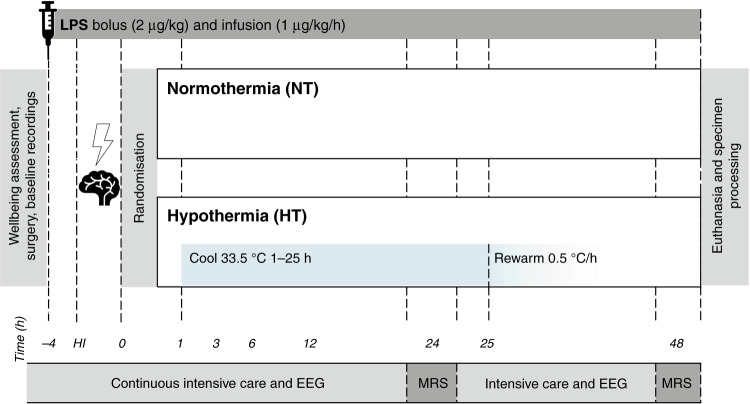
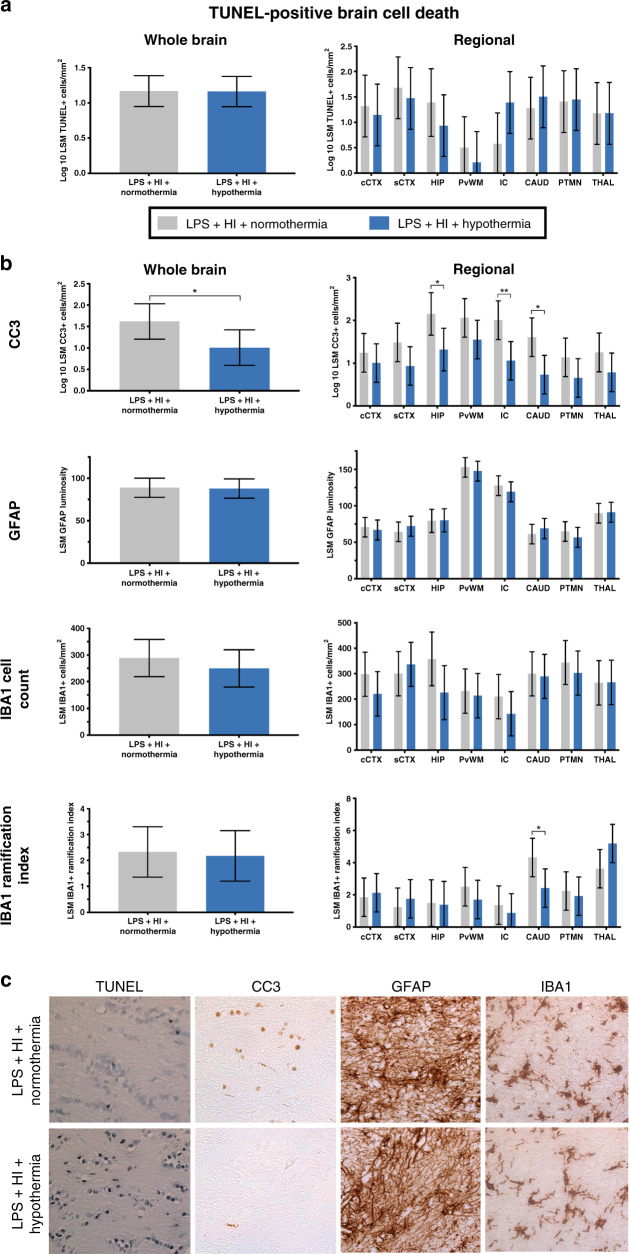
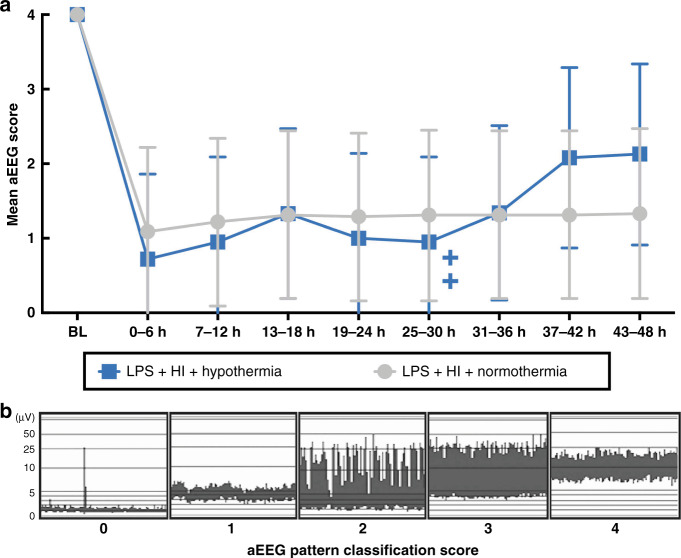
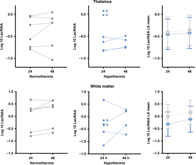
Methods: Twelve newborn piglets received a 2 µg/kg bolus and 1 µg/kg/h infusion over 52 h of Escherichia coli lipopolysaccharide (LPS). HI was induced 4 h after LPS bolus. After HI, piglets were randomized to HT (33.5 °C 1-25 h after HI, n = 6) or normothermia (NT, n = 6). Amplitude-integrated electroencephalogram (aEEG) was recorded and magnetic resonance spectroscopy (MRS) was acquired at 24 and 48 h. At 48 h, terminal deoxynucleotidyl transferase dUTP nick-end labeling (TUNEL)-positive brain cell death, microglial activation/proliferation, astrogliosis, and cleaved caspase-3 (CC3) were quantified. Hematology and plasma cytokines were serially measured.

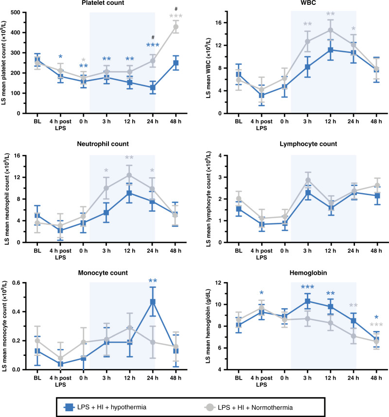
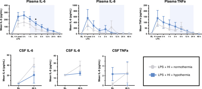
Results: Two HT piglets died. aEEG recovery, thalamic and white matter MRS lactate/N-acetylaspartate, and TUNEL-positive cell death were similar between groups. HT increased microglial activation in the caudate, but had no other effect on glial activation/proliferation. HT reduced CC3 overall. HT suppressed platelet count and attenuated leukocytosis. Cytokine profile was unchanged by HT.

Conclusions: We did not observe protection with HT in this piglet IS-HI model based on aEEG, MRS, and immunohistochemistry. Immunosuppressive effects of HT and countering neuroinflammation by LPS may contribute to the observed lack of HT efficacy. Other immunomodulatory strategies may be more effective in IS-HI.

Impact: Acute infection/inflammation is known to exacerbate perinatal brain injury and can worsen the outcomes in neonatal encephalopathy. Therapeutic HT is the current standard of care for all infants with NE, but the benefit in infants with coinfection/inflammation is unknown. In a piglet model of inflammation (LPS)-sensitized HI, we observed no evidence of neuroprotection with cooling for 24 h, based on our primary outcome measures: aEEG, MRS Lac/NAA, and histological brain cell death. Additional neuroprotective agents, with beneficial immunomodulatory effects, require exploration in IS-HI models.


Conflict of interest statement

The authors declare no competing interests.

Figures

Fig. 1
Fig. 1. Study timeline.
Following baseline recordings and surgery, piglets received a 2 µg/kg LPS bolus, followed by 1 µg/kg/h LPS infusion continued for 52 h. After 4 h of infusion, piglets underwent a transient hypoxic–ischemic (HI) insult. Animals were subsequently randomized to (i) normothermia (NT, core 38.0–39.0 °C throughout) or (ii) hypothermia (HT, 33.5 °C from 1 to 25 h post HI, then rewarmed at 0.5 °C/h). Blood samples were collected at baseline, 4 h after bolus, end HI (time 0), and at 3, 12, 24, and 48 h after HI. EEG was continuously acquired, excluding during MRS at 24 and 48 h. Piglets were maintained under meticulous intensive care for 48 h following HI, prior to euthanasia and histological specimen collection.
Fig. 2
Fig. 2. Brain histology.
a TUNEL histology. There was no difference between whole-brain or regional estimated least-squares mean (LSM) TUNEL-positive cells per mm2. b LSM whole-brain and regional cleaved caspase-3 (CC3), GFAP, IBA1-positive cell count, and IBA1 ramification index. Hypothermia reduced CC3 throughout the brain and in the internal capsule, caudate nucleus, and hippocampus. Hypothermia did not alter astrogliosis. Cooling had no effect on the overall microglial number or activation state. There was an increase in microglial activation (lower ramification index) in the caudate with HT. c Representative sections for each stain (TUNEL, CC3, GFAP, and IBA1) from the internal capsule are shown at ×40 magnification. cCTX cingulate cortex, sCTX sensorimotor cortex, HIP hippocampus, PvWM periventricular white matter, IC internal capsule, CAUD caudate, PTMN putamen, THAL thalamus. Data are displayed as analyzed, on a log 10 scale. Error bars represent 95% CI. *p < 0.05, **p < 0.01.
Fig. 3
Fig. 3. aEEG.
Mean aEEG pattern classification score from baseline (BL) till 48 h, divided into 6 h time epochs (a) (±95% CI). There was no difference between NT and HT aEEG scores at any time. The two crosses represent the deaths of two HT piglets at 30 and 33 h. Examples of each pattern classification score are shown (b), where 0 = flat trace, 1 = low voltage, 2 = burst suppression, 3 = discontinuous normal voltage, and 4 = continuous normal voltage.
Fig. 4
Fig. 4. MRS.
There is no effect of hypothermia on the ratio of lactate to N-acetylaspartate (Lac/NAA) in the thalamus or white matter at 24 or 48 h after HI. In all subjects, log 10 Lac/NAA data points are shown in the graphs on the left/center, and are summarized using log 10 least-squares (LS) means plot with 95% CI error bars in the boxes on the right. The crosses indicate the two HT piglets who died prior to the 48 h scan.
Fig. 5
Fig. 5. Hematology.
Least-squares mean values over time for platelets, white blood cells (WBC), neutrophils, lymphocytes, monocyte counts, and hemoglobin (±SEM). Pale blue shading represents cooling. *P < 0.05, **p < 0.01, and ***p < 0.0001 for change from baseline value, with color denoting group; #p < 0.05 for the difference at timepoint between the NT and HT group.
Fig. 6
Fig. 6. Cytokines.
Mean values over time for IL-6, IL-8, and TNFα in the plasma and cerebrospinal fluid (CSF) (±SEM). Pale blue shading represents cooling. *P < 0.05 for difference at timepoint between the NT and HT group.

References

    1. Jacobs, S. E. et al. Cooling for newborns with hypoxic ischaemic encephalopathy. Cochrane Database of Systematic Reviews, CD003311. 10.1002/14651858.CD003311.pub3 (2013). - PMC - PubMed
    1. Shankaran S, et al. Childhood outcomes after hypothermia for neonatal encephalopathy. N. Engl. J. Med. 2012;366:2085–2092. doi: 10.1056/NEJMoa1112066. - DOI - PMC - PubMed
    1. Jary S, Smit E, Liu X, Cowan FM, Thoresen M. Less severe cerebral palsy outcomes in infants treated with therapeutic hypothermia. Acta Paediatr. 2015;104:1241–1247. doi: 10.1111/apa.13146. - DOI - PubMed
    1. Lee-Kelland R, et al. School-age outcomes of children without cerebral palsy cooled for neonatal hypoxic-ischaemic encephalopathy in 2008-2010. Arch. Dis. Child. Fetal Neonatal Ed. 2020;105:8–13. doi: 10.1136/archdischild-2018-316509. - DOI - PubMed
    1. Badawi N, et al. Antepartum risk factors for newborn encephalopathy: the Western Australian case-control study. BMJ. 1998;317:1549–1553. doi: 10.1136/bmj.317.7172.1549. - DOI - PMC - PubMed

Publication types

Substances