. 2021 Oct;21(10):3239-3255.
doi: 10.1111/ajt.16611. Epub 2021 Jun 24.

Recipient sex and estradiol levels affect transplant outcomes in an age-specific fashion

Affiliations

Recipient sex and estradiol levels affect transplant outcomes in an age-specific fashion

Ryoichi Maenosono et al. Am J Transplant. 2021 Oct.

Abstract

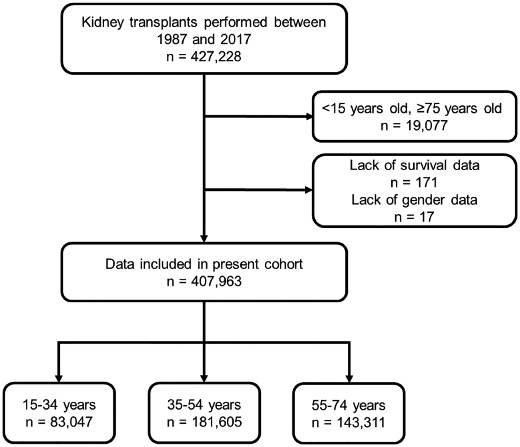
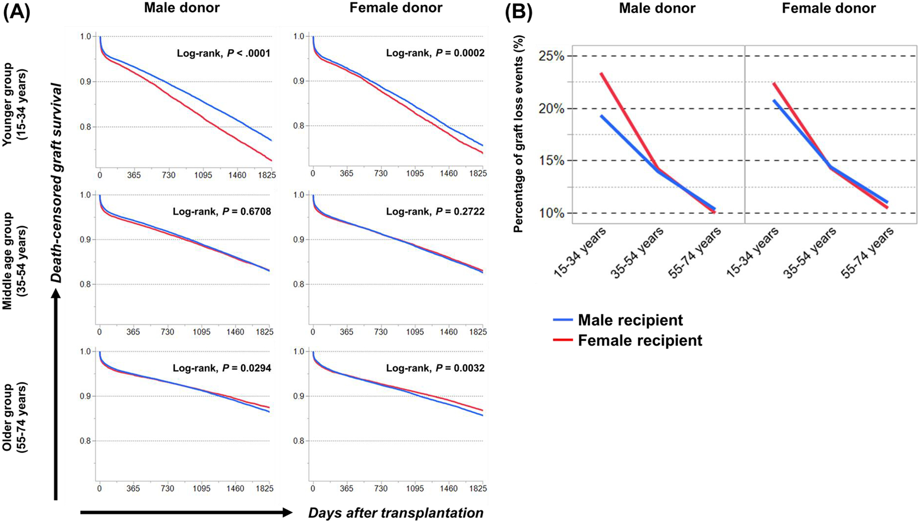
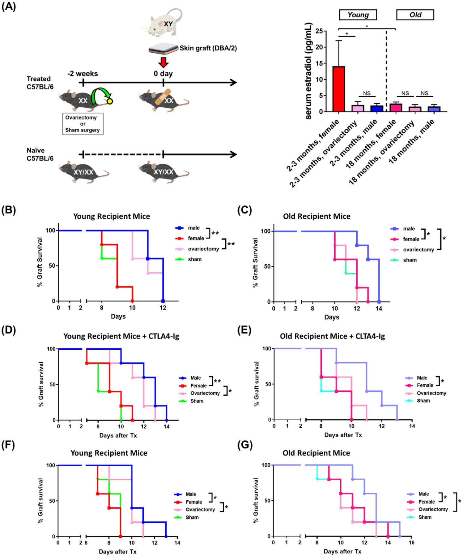
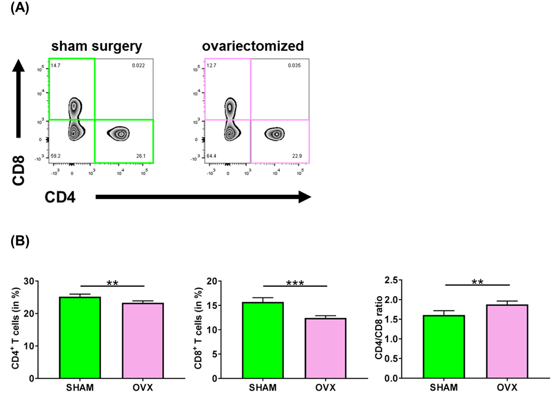
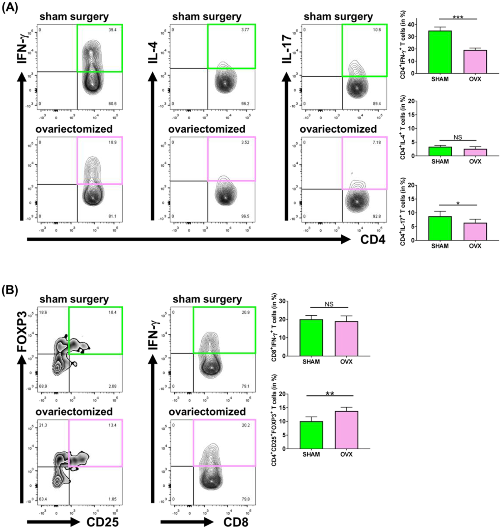
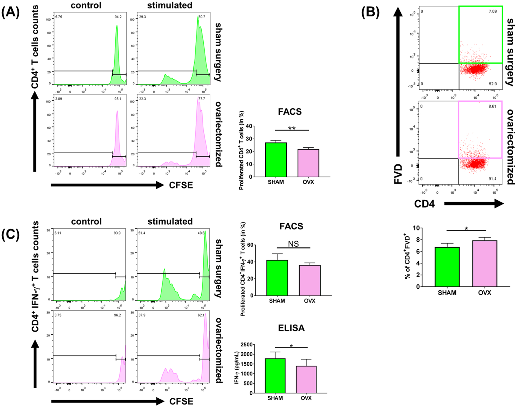
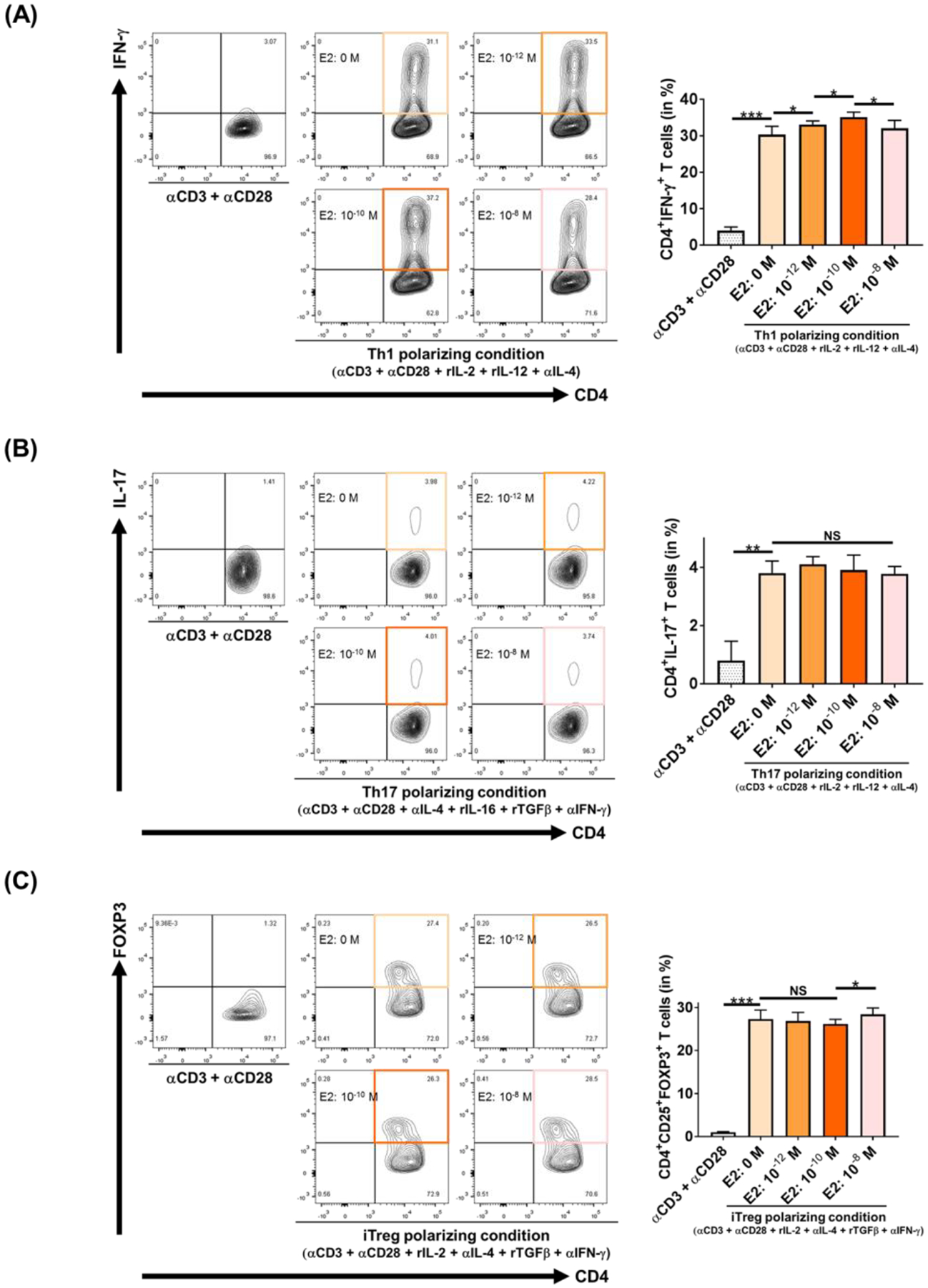
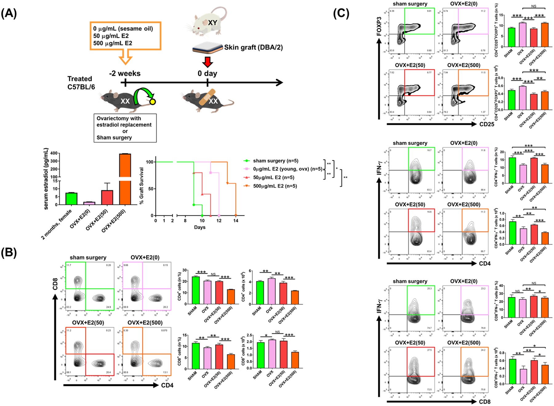
Sex-specific influences have been shown for a variety of diseases. Whether donor or recipient sex and sex hormone levels impact alloimmune responses remains unclear. In unifactorial and multifactorial analyses of more than 400 000 SRTR listed kidney transplant patients, we found that younger female recipients had an inferior death-censored graft survival that was independent of donor sex. In contrast, graft survival was superior in older female recipients, suggesting the impact of recipient sex hormones over chromosomal sex mismatches. Those clinical changes were delineated in experimental skin and heart transplant models showing a prolongation of graft survival in ovariectomized young female recipients. In contrast, graft survival was comparable in ovariectomized and naïve old female recipients. Young ovariectomized mice showed reduced amounts and a compromised T cell proliferation. Deprivation of female hormones dampened the production of interferon (IFN)-γ and interleukin (IL)-17+ by CD4+ T cells while augmenting systemic counts of Tregs. Increasing estradiol concentrations in vitro promoted the switch of naïve CD4+ T cells into Th1 cells; high physiological estradiol concentrations dampening Th1 responses, promoted Tregs, and prolonged graft survival. Thus, clinical observations demonstrate age-specific graft survival patterns in female recipients. Estrogen levels, in turn, impact the fate of T cell subsets, providing relevant and novel information on age- and sex-specific alloimmunity.

Keywords: basic (laboratory) research/science; endocrinology/diabetology; gender; kidney failure/injury; kidney transplantation/nephrology; organ allocation; organ transplantation in general; rejection: T cell mediated (TCMR); reproductive biology.


Conflict of interest statement

Conflict of interest

The authors declare no conflict of interest.

Figures

Figure 1.
Figure 1.
Out of a total of 427,228 recipients transplanted between 1987 – 2017, we excluded 19,077 (<15 years or ≥75 years); 188 patients were excluded with insufficient data. The remaining 407,963 cases were separated into three groups according to younger (15–34 years, n = 83,047), middle age (35–54 years, n = 181,605) or older (55–75 years, n = 143,311).
Figure 2.
Figure 2.
Both, donor sex and recipients age impacted death-censored graft survivals outcome. (A) Kaplan-Meier curves showing the impact of recipients age and donor sex on 5 years death-censored graft survival outcome. (B) Percentage of graft loss/recipient age (15–34, 35–54 and 55–74 years) and donor sex; P values were calculated using a two-sided log rank test. Blue lines represent male and red line female recipients; *P < 0.05; **P < 0.01; ***P < 0.0001).
Figure 3.
Figure 3.
Survival of fully mismatched skin allografts after ovariectomies are shown. (A) Model: C57BL/6 underwent ovariectomies or sham surgeries prior to skin transplantation; after two weeks, mice received a fully mismatch skin transplant. Skin allografts from young male DBA/2 mice were transplanted onto young (2–3 months) or old C57BL/6 mice (18 months). (B) Skin survival of naïve young male and female recipients in addition to female animals that underwent ovariectomies or sham surgeries are shown (n = 5 per group). (C) Skin survival of old naïve male and female recipients, old females that underwent ovariectomies or sham surgeries are shown (n = 5/group). (D) (E) Survival after skin transplants onto young and old naïve male, naïve female, ovariectomized female and sham operated recipients treated with either CTLA4-Ig or PBS are shown. (F) and (G) cardia allograft survival in young and old naïve male, naïve female, ovariectomized female and sham operated recipients are shown. Log-rank test was used to compare graft survival; *P < 0.05; **P < 0.01; ***P < 0.0001
Figure 4.
Figure 4.
CD4+ and CD8+ T cell frequencies of young skin transplanted animals after ovariectomies or sham surgeries are shown. Eight days after skin transplantation and prior to rejection, spleens were collected, and single leukocytes suspensions were obtained. Frequencies of CD4+ and CD8+ T cells were assessed using flow cytometry (n = 5 per group; data represent mean ± SD; data were compared by applying Student t test (*P < 0.05; **P < 0.01; ***P < 0.0001).
Figure 5.
Figure 5.
Ovariectomies modified alloimmune responses in young female recipients. Young (2–3 months) C57BL/6 female mice underwent ovariectomies or sham surgeries and received fully mismatch skin transplants from male donors. By day 8, prior to skin transplantation, spleens were collected, and single leukocytes suspensions were obtained. Frequencies of CD4+IFN-γ+ (Th1), CD4+IL-4+ (Th2), CD4+IL-17+ (Th17), CD4+CD25+FOXP3+ (Treg) and CD8+IFN-γ+ cells were assessed by flow cytometry (n = 5 per group; data represent mean ± SD; The Student t test was used to compare groups; *P < 0.05; **P < 0.01; ***P < 0.0001).
Figure 6.
Figure 6.
Ovariectomies reduced T cell proliferation and promoted T cell death in young female recipients. Young (2–3 months) C57BL/6 female mice underwent ovariectomies or sham surgeries and received fully mismatch skin transplant (DBA/2 young (2–3 month) male donors). Eight days after skin transplantation (a day prior full rejection), spleens were collected, and single leukocytes suspensions were obtained. To determine the proliferation of CD4+ T and CD4+IFN-γ+ cells, splenocytes were stained with CFSE dilution and cultured with stimulator DBA/2 splenocytes (MLR) treated with mitomycin. After 5 days of culture, cells were collected and analyzed by flow cytometry (n = 5 per group; data represent mean ± SD; Student t test was used to compare groups; *P < 0.05; **P < 0.01; ***P < 0.0001).
Figure 7.
Figure 7.
17β-estradiol modifies IFN-γ+ production and CD4+CD25+FOXP3+ regulatory T cells under Th1 and iTreg polarizing conditions. Single cell suspensions were obtained from spleens of young C57BL/6 mice; CD4+ T cells were isolated by negative selection (purity > 95%). CD4+ T cells were then cultured under (A) Th1, (B) Th17 or (C) iTreg polarizing conditions with differing concentrations of 17β-estradiol. By 5 days, cells were stained for double stained for CD4, CD25 and the intracellular expression of IFN-γ, IL-17 and FOXP3 (n = 5 per group; data represent mean ± SD; Student t test and ANOVA were used to compare groups; *P < 0.05; **P < 0.01; ***P < 0.0001).
Figure 8.
Figure 8.
Hormone replacement reverses the effects of ovariectomies on alloimmune responses and transplant survival. (A) Young female C57BL/6 mice (2–3 months) underwent sham or estradiol treatment prior to skin transplantation. Treatments changed mice serum estradiol level. Two weeks later, mice received fully mismatched skin transplants. Skin allografts from young male DBA/2 mice were transplanted onto young female recipient mice (2–3 months). (B) Prior to rejection, spleens were collected, and single leukocytes suspensions were obtained. Frequencies of young CD4+ and CD8+ T cells (2–3 months) were assessed by flow cytometry (n = 5 animals/group). (C) Frequencies of CD4+CD25+FOXP3+ Tregs, CD4+IFN-γ+ and CD8+IFN-γ+ T cells were assessed by flow cytometry (n = 5 per group; data represent mean ± SD; Student t test and ANOVA were used to compare groups; *P < 0.05; **P < 0.01; ***P < 0.0001).

Comment in

Similar articles

Cited by

References

    1. Ngo ST, Steyn FJ, McCombe PA. Gender differences in autoimmune disease. Front Neuroendocrinol 2014;35(3):347–369. - PubMed
    1. Regitz-Zagrosek V, Kararigas G. Mechanistic Pathways of Sex Differences in Cardiovascular Disease. Physiol Rev 2017;97(1):1–37. - PubMed
    1. Pike CJ. Sex and the development of Alzheimer’s disease. J Neurosci Res 2017;95(1–2):671–680. - PMC - PubMed
    1. McClelland EE, Smith JM. Gender specific differences in the immune response to infection. Arch Immunol Ther Exp (Warsz) 2011;59(3):203–213. - PubMed
    1. Gebhard C, Regitz-Zagrosek V, Neuhauser HK, Morgan R, Klein SL. Impact of sex and gender on COVID-19 outcomes in Europe. Biol Sex Differ 2020;11(1):29. - PMC - PubMed

Publication types