

Immune responses to injury and their links to eye disease
- PMID: 34051364
- PMCID: PMC8380715
- DOI: 10.1016/j.trsl.2021.05.005
Immune responses to injury and their links to eye disease
Abstract
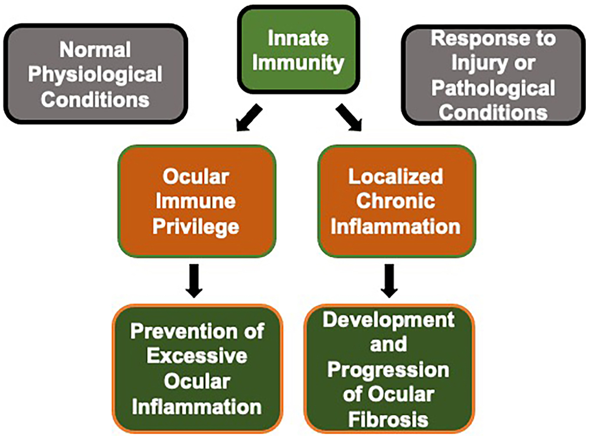
The eye is regarded as an immune privileged site. Since the presence of a vasculature would impair vision, the vasculature of the eye is located outside of the central light path. As a result, many regions of the eye evolved mechanisms to deliver immune cells to sites of dysgenesis, injury, or in response to the many age-related pathologies. While the purpose of these immune responses is reparative or protective, cytokines released by immune cells compromise visual acuity by inducing inflammation and fibrosis. The response to traumatic or pathological injury is distinct in different regions of the eye. Age-related diseases impact both the anterior and posterior segment and lead to reduced quality of life and blindness. Here we focus attention on the role that inflammation and fibrosis play in the progression of age-related pathologies of the cornea and the lens as well as in glaucoma, the formation of epiretinal membranes, and in proliferative vitreoretinopathy.
Keywords: 2ryERM; A T-helper cell that expresses high levels of IL-17 which can suppress T-regulatory cell function; A cytokine expressed early during inflammation that attracts neutrophils; A cytokine expressed early during inflammation that attracts neutrophils, sometimes referred to as monocyte chemoattractant protein-1 (MCP-1)); A mouse model that lacks functional T and B cells and used to study the immune response; A pigmented mouse strain used for research and known to mount a primarily Th1 response to infection; A protein encoded by the ADGRE1 gene that, in mice, is expressed primarily on macrophages; A strain of pigmented mice used in glaucoma research; ACAID; APCs; ASC; An albino mouse strain used for research and known to mount a primarily Th2 response to infection; Antigen Presenting Cells, this class includes dendritic cells and monocytes; BALB/c; BM; C57BL6; CCL2; CD45; CNS; CXCL1; Central Nervous System; Cluster of differentiation 45 antigen; DAMPs; DBA/2J; EBM; ECM; EMT; ERM; Epithelial Basement Membrane; F4/80; FGF2; HA =hyaluronic acid; HSK; HSP; HSPGs; HSV; ICN; IL-20; IL6; ILM; IOP; Inner (or internal) limiting membrane; Interleukin 6; Interleukin-20; MAGP1; MHC-II; Major histocompatibility complex type II, a class of MHC proteins typically found only on APCs; Microfibril-associated glycoprotein 1; N-cad; N-cadherin; NEI; NK; National Eye Institute; Natural killer T cells; PCO; PDGF; PDR; PVD; PVR; Platelet derived growth factor; Posterior capsular opacification; RGC; RPE; RRD; Rag1-/-; Retinal ganglion cells; Retinal pigment epithelial cells; SMAD; Sons of Mothers Against Decapentaplegic, SMADs are a class of molecules that mediate TGF and bone morphogenetic protein signaling; T-helper cell 1 response, proinflammatory adaptive response involving interferon gamma and associated with autoimmunity; T-helper cell 2 response involving IgE and interleukins 4,5, and 13, also induces the anti-inflammatory interleukin 10 family cytokines; T-regulatory cell; TG; TGF1; TM; TNF; Th1; Th17; Th2; Transforming growth factor 1; Treg; Tumor necrosis factor a cytokine produced during inflammation; VEGF; Vascular endothelial growth factor; WHO; World Health Organization; anterior chamber immune deviation; anterior subcapsular cataracts; basement membrane; damage-associated molecular patterns; epiretinal membrane; epiretinal membrane secondary to disease pathology; epithelial-mesenchymal transition; extracellular matrix; fibroblast growth factor 2, also referred to as basic FGF; heat shock protein; heparan sulfate proteoglycans; herpes simplex virus; herpes stromal keratitis; iERM; idiopathic epiretinal membrane; intraepithelial corneal nerves; intraocular pressure; mTOR; mechanistic target of rapamycin, a protein kinase encoded by the MTOR genes that regulates a variety of signal transduction events including cell growth, autophagy and actin cytoskeleton; posterior vitreous detachment; proliferative diabetic retinopathy; proliferative vitreoretinopathy; rhegmatogenous (rupture, tear) retinal detachment; trabecular meshwork; trigeminal ganglion; αSMA; α−Smooth muscle actin, a class of actin expressed in mesenchymal cells.
Copyright © 2021 The Author(s). Published by Elsevier Inc. All rights reserved.
Figures


References
-
- Friend J, Thoft RA. Functional competence of regenerating ocular surface epithelium. Invest Ophthalmol Vis Sci. 1978;17:134–9. - PubMed
-
- Thoft RA. Role of the ocular surface in destructive corneal disease. Trans Ophthalmol Soc U K. 1978;98:339–42. - PubMed
-
- Thoft RA, Friend J, Kenyon KR. Ocular surface response to trauma. Int Ophthalmol Clin. 1979;19:111–31. - PubMed
-
- Hamrah P, Zhang Q, Liu Y, Dana MR. Novel characterization of MHC class II-negative population of resident corneal Langerhans cell-type dendritic cells. Invest Ophthalmol Vis Sci. 2002;43:639–46. - PubMed
-
- Hamrah P, Liu Y, Zhang Q, Dana MR. The corneal stroma is endowed with a significant number of resident dendritic cells. Invest Ophthalmol Vis Sci. 2003;44:581–9. - PubMed
Publication types
MeSH terms
Grants and funding
LinkOut - more resources
Full Text Sources
Other Literature Sources
Medical
Research Materials
Miscellaneous
