Review
. 2021 Jul:121:102662.
doi: 10.1016/j.jaut.2021.102662. Epub 2021 May 19.

Mechanisms of Immunothrombosis in Vaccine-Induced Thrombotic Thrombocytopenia (VITT) Compared to Natural SARS-CoV-2 Infection

Affiliations
Review

Mechanisms of Immunothrombosis in Vaccine-Induced Thrombotic Thrombocytopenia (VITT) Compared to Natural SARS-CoV-2 Infection

Dennis McGonagle et al. J Autoimmun. 2021 Jul.

Abstract

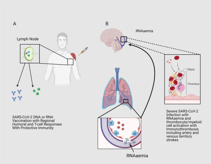
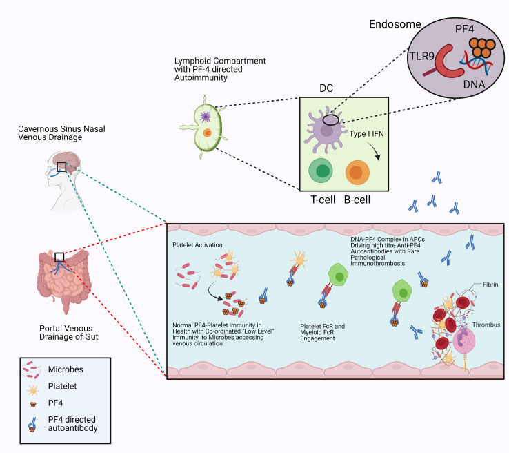
Herein, we consider venous immunothrombotic mechanisms in SARS-CoV-2 infection and anti-SARS-CoV-2 DNA vaccination. Primary SARS-CoV-2 infection with systemic viral RNA release (RNAaemia) contributes to innate immune coagulation cascade activation, with both pulmonary and systemic immunothrombosis - including venous territory strokes. However, anti-SARS-CoV-2 adenoviral-vectored-DNA vaccines -initially shown for the ChAdOx1 vaccine-may rarely exhibit autoimmunity with autoantibodies to Platelet Factor-4 (PF4) that is termed Vaccine-Induced Thrombotic Thrombocytopenia (VITT), an entity pathophysiologically similar to Heparin-Induced Thrombocytopenia (HIT). The PF4 autoantigen is a polyanion molecule capable of independent interactions with negatively charged bacterial cellular wall, heparin and DNA molecules, thus linking intravascular innate immunity to both bacterial cell walls and pathogen-derived DNA. Crucially, negatively charged extracellular DNA is a powerful adjuvant that can break tolerance to positively charged nuclear histone proteins in many experimental autoimmunity settings, including SLE and scleroderma. Analogous to DNA-histone interactons, positively charged PF4-DNA complexes stimulate strong interferon responses via Toll-Like Receptor (TLR) 9 engagement. A chain of events following intramuscular adenoviral-vectored-DNA vaccine inoculation including microvascular damage; microbleeding and platelet activation with PF4 release, adenovirus cargo dispersement with DNA-PF4 engagement may rarely break immune tolerance, leading to rare PF4-directed autoimmunity. The VITT cavernous sinus cerebral and intestinal venous territory immunothrombosis proclivity may pertain to venous drainage of shared microbiotal-rich areas of the nose and in intestines that initiates local endovascular venous immunity by PF4/microbiotal engagement with PF4 autoantibody driven immunothrombosis reminiscent of HIT. According to the proposed model, any adenovirus-vectored-DNA vaccine could drive autoimmune VITT in susceptible individuals and alternative mechanism based on molecular mimicry, vaccine protein contaminants, adenovirus vector proteins, EDTA buffers or immunity against the viral spike protein are secondary factors. Hence, electrochemical DNA-PF4 interactions and PF4-heparin interactions, but at different locations, represent the common denominator in HIT and VITT related autoimmune-mediated thrombosis.

Keywords: COVID-19 pneumonia related thrombosis; Heparin induced thrombocytopenia (HIT) DNA-PF4 interactions. VITT model; Vaccine induced thrombotic thrombocytopenia (VITT).


Figures

Fig. 1
Fig. 1
Immunothrombosis in natural SARS-CoV-2 infection and vaccine prevention. A) RNA and DNA SARS-CoV-2 vaccines are not associated with viral replication and systemic RNAaemia or pulmonary vascular territory immunothrombosis as a small nucleic acid inoculums are confined to the muscle and are therefore not linked to innate immune mediated thrombosis. B) Natural SARS-CoV-2 infection is associated with extensive pulmonary capillary, pulmonary arteriolar and pulmonary venular territory immunothrombosis in severe COVID-19. Viral RNA is a potent activation of the coagulation cascade and likely contributes to the immunothrombosis phenotype including arterial and venous territory strokes. Additionally, severe COVID-19 pneumonia appears to be associated with alveolar-capillary barrier breakdown with access of SARS-CoV-2 RNA or RNAaemia which can trigger an innate immune driven venous thrombosis in multiple organs including the brain. So RNAaemia and hypercoagulability from innate immune cell activation within the lung and other factors in severe COVID-19 pneumonia may predispose to venous territory strokes. Although HIT may also exhibit arterial and venous strokes it has a distinct autoimmune component.
Fig. 2
Fig. 2
Vascular and immune topography of VITT The VITT associated thrombosis is predominantly venous and involves the gastrointestinal portal circulation and the cavernous cerebral venous sinus circulation. Both of these venous territories share the common feature of draining the nasal sinus and intestines, thus allowing access of microbiotal, viral products and potentially toxins to the endothelial networks of lining vessels. Microbe platelet interactons with platelet factor-4 (PF4) coating and PF4/PF4 “natural” autoantibody cross-talk with platelets and myeloid cells maintains immunity and homeostasis restoration. The presence on high titre autoantibodies against PF4 in these tissue specific sites of susceptibility may lead to an exaggerated immune response including platelet and neutrophil activation, increased platelet consumption and thrombosis. Sequestration of PF4 in the presence of anti-PF4 autoantibodies triggers extensive FcRγII mediated platelet activation with further PF4 release and further platelet and immune cell chemotaxis thus initiating systemic thrombosis and platelet consumption which is based on the model of PF4 immune activation in HIT. The interaction between highly negatively charged heparin molecules on endothelial surfaces and the highly positively charged PF4 in the clot environment could further increase immunothrombosis.

References

    1. Cines D.B., Bussel J.B. SARS-CoV-2 vaccine-induced immune thrombotic thrombocytopenia. N. Engl. J. Med. 2021 doi: 10.1056/NEJMe2106315. - DOI - PMC - PubMed
    1. Schultz N.H., et al. Thrombosis and thrombocytopenia after ChAdOx1 nCoV-19 vaccination. N. Engl. J. Med. 2021 doi: 10.1056/NEJMoa2104882. - DOI - PMC - PubMed
    1. Mehta P.R., et al. Cerebral venous sinus thrombosis and thrombocytopenia after COVID-19 vaccination - a report of two UK cases. Brain Behav. Immun. 2021 doi: 10.1016/j.bbi.2021.04.006. - DOI - PMC - PubMed
    1. McGonagle D., et al. COVID-19 vasculitis and novel vasculitis mimics. Lanc Rheumatol. 2021;3(3):e224–e233. - PMC - PubMed
    1. Harrison S.R., et al. Chest pain mimicking pulmonary embolism may be a common presentation of COVID-19 in ambulant patients without other typical features of infection. J. Intern. Med. 2021 doi: 10.1111/joim.13267. - DOI - PMC - PubMed

MeSH terms