. 2021 Jul:274:120874.
doi: 10.1016/j.biomaterials.2021.120874. Epub 2021 May 12.

Biomimetic tubular scaffold with heparin conjugation for rapid degradation in in situ regeneration of a small diameter neoartery

Affiliations

Biomimetic tubular scaffold with heparin conjugation for rapid degradation in in situ regeneration of a small diameter neoartery

Renato S Navarro et al. Biomaterials. 2021 Jul.

Abstract

To address the clinical need for readily available small diameter vascular grafts, biomimetic tubular scaffolds were developed for rapid in situ blood vessel regeneration. The tubular scaffolds were designed to have an inner layer that is porous, interconnected, and with a nanofibrous architecture, which provided an excellent microenvironment for host cell invasion and proliferation. Through the synthesis of poly(spirolactic-co-lactic acid) (PSLA), a highly functional polymer with a norbornene substituting a methyl group in poly(l-lactic acid) (PLLA), we were able to covalently attach biomolecules onto the polymer backbone via thiol-ene click chemistry to impart desirable functionalities to the tubular scaffolds. Specifically, heparin was conjugated on the scaffolds in order to prevent thrombosis when implanted in situ. By controlling the amount of covalently attached heparin we were able to modulate the physical properties of the tubular scaffold, resulting in tunable wettability and degradation rate while retaining the porous and nanofibrous morphology. The scaffolds were successfully tested as rat abdominal aortic replacements. Patency and viability were confirmed through dynamic ultrasound and histological analysis of the regenerated tissue. The harvested tissue showed excellent vascular cellular infiltration, proliferation, and migration with laminar cellular arrangement. Furthermore, we achieved both complete reendothelialization of the vessel lumen and native-like media extracellular matrix. No signs of aneurysm or hyperplasia were observed after 3 months of vessel replacement. Taken together, we have developed an effective vascular graft able to generate small diameter blood vessels that can function in a rat model.

Keywords: Heparin conjugation; Small diameter vascular graft; Tubular scaffold; Vascular smooth muscle cells; Vascular tissue engineering.


Conflict of interest statement

Declaration of interests

The authors declare that they have no known competing financial interests or personal relationships that could have appeared to influence the work reported in this paper.

Figures

Figure 1.
Figure 1.
Characterization of PSLA and precursor monomers. 1A-C) 500 MHz 1H NMR spectrum of exomethylene lactide (1A), spirolactide (1B), 1:3 poly(spirolactic-co-lactic acid) (1C) in CDCl3. 1D-G) FTIR characterization depicting stretches of key functional groups of L-lactide (1D), exomethylene lactide (1E), spirolactide (1F), and PSLA (1G) to compare absorption peaks. 1H) UV – Vis absorbance of monomers (PLLA, methylene lactide, spirolactide) and resulting polymers (PLLA and PSLA) to compare their absorption peaks and trends.
Figure 2.
Figure 2.
Fabrication of a tubular scaffold that can be easily post-modified. A) Fabrication method of sugar template annealing on a mandrel to fabricate highly porous and interconnected pore network. B) Overview and SEM characterization of PSLA/PLLA loose layer that is highly porous, interconnected, and nanofibrous. C) SEM visualization of PCL dense layer that is electrospun to provide enhanced mechanical support. Green box = magnified image of fiber morphology, yellow box = interface between the outer PCL layer and a portion of the PSLA/PLLA “loose layer” (inner porous scaffold).
Figure 3.
Figure 3.
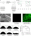
Post-modification via thiol-ene click chemistry and characterization. A) Post-modification scheme of PSLA polymer with heparin via thiol-ene click chemistry. B) FTIR spectra demonstrating pre- and post-modification structural changes. C) SEM visualization of tubular scaffold post modification. D) Confocal image of PLLA and PSLA modified with FITC-PEG-SH fluorescent probe. E) Change of hydrophilicity observation, quantification, and comparison of PLLA to PLLA/PSLA, PLLA w/ heparin, and PLLA/PSLA with increasing concentrations of heparin: not significant (ns), ns, and significant with **** p < 0.0001, respectively.
Figure 4.
Figure 4.
Degradation and mass loss of PLLA/PSLA scaffolds after heparin conjugation. A) Visualization of PSLA scaffold conjugated with heparin began to degrade by week 2 compared to all other groups that remained intact at the end of the experiment with minimal degradation, where the scaffold dimensions were 22 mm in diameter and 2.0 mm in thickness. B) Quantification of scaffold mass loss over time and observed statistical significance of PLLA compared to PLLA with heparin addition, PLLA/PSLA, and heparin conjugated PLLA/PSLA scaffolds: not significant (ns), ns, and statistically significant with ****p < 0.0001, respectively. C) SEM characterization of scaffold degradation at day 0 and day 35, scale bar 100 μm and 20 μm (insets)
Figure 5.
Figure 5.
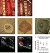
Scaffold evaluation post-implantation in situ. A – B) Operative images of implanted scaffolds immediately after implantation (A) and 3 months post-operation (B). C – E) Morphologies of nanofibrous vascular scaffolds before rat abdominal aortic interpositional implantation (C), 3 months post-operation (D), and native rat abdominal aorta (E). F & G) Ultrasound images of an implanted scaffold 3 months post-operation. H) Comparison of native aorta vs TEBVs over 3-month period: 1 month (n = 24), 2 months (n = 15), 3 months (n = 12), p > 0.05.
Figure 6.
Figure 6.
Comparison of vascular muscular reconstruction at anastomosis and middle sites of implanted scaffolds using H&E staining. Reconstruction of the anastomosis site of scaffold at 1 week (A, B), 2 weeks (E, F), 1 month (I, J), and 3 months post-operation (M, N). Reconstruction of the middle site of scaffold at 1 week (C, D), 2 weeks (G, H), 1 month (K, L), 3 months post-operation (O, P), and rat native aorta (Q, R). (Scale bar: A, C, E, G, I, K, M, O, Q = 400 μm; B, D, F, H, J, L, N, P, R = 40 μm).
Figure 7.
Figure 7.
Comparison of vascular extracellular matrix reconstruction of implanted scaffolds after 1 and 3 months of implantation. Analysis of collagen by Masson Trichrome staining of rat native aorta (A, B), implanted scaffold at 1 month (E, F), and 3 months post-op (I, J). Analysis of elastin by Verhoeff Van Gieson staining of rat native aorta (C, D), implanted scaffold at 1 month post-op (G, H), and 3 months post-op (K, L). Scale bars: A, C, E, G, I, K = 400 μm; B, D, F, H, J, L = 40 μm.
Figure 8A.
Figure 8A.
Immunofluorescent staining for smooth muscle cell marker SM22 indicates smooth muscle cells infiltration into scaffolds and vessel reconstruction in rat aorta, 1 month post-op and 3 month post-op in scaffolds. Immuno-fluorescent staining for SM22, nuclear staining, and merged images in native rat aorta (A, D, G), implanted scaffold at 1 month (B, E, H), and implanted scaffold at 3 months post-op (C, F, I). The scale bar is equal to 40 μm.
Figure 8B.
Figure 8B.
Immunofluorescent staining of smooth muscle cell contractile protein marker MYH11 indicates smooth muscle cell infiltration and vessel reconstruction in rat aorta at 1 month and 3-months post-op of tubular scaffolds. Immunofluorescent staining of MYH11, nuclear staining, and merged images in native rat aorta (A, D, G), implanted scaffold at 1 month (B, E, H), and implanted scaffold at 3 months post-op (C, F, I). The scale bar is equal to 40 μm.
Figure 9.
Figure 9.
Immunohistochemical staining of endothelial marker vWF of implanted scaffolds indicates endothelial reconstruction. Native rat aorta (A), Implanted scaffold for 2 weeks (B), 1 month (C), and 3 months post-op (D). (Scale bar = 40um).
Scheme 1.
Scheme 1.
Synthesis route of PLLA-based copolymer poly(spirolactic-co-lactic acid). A) Addition of bromine from NBS followed by the elimination of the bromine using TEA to produce exomethylene lactide. B) Diels-Alder addition of freshly distilled cyclopentadiene refluxed with exomethylene lactide to yield spirolactide. C) Polymerization of PSLA from L-lactide and spirolactide.

References

    1. Matheny M; McPheeters ML; Glasser A; Mercaldo N; Weaver RB; Jerome RN; Walden R; McKoy JN; Pritchett J; Tsai C, In Systematic Review of Cardiovascular Disease Risk Assessment Tools, Rockville (MD), 2011. - PubMed
    1. Kaplan GA; Keil JE, Socioeconomic factors and cardiovascular disease: a review of the literature. Circulation 1993, 88 (4 Pt 1), 1973–98. - PubMed
    1. In Health, United States, 2010: With Special Feature on Death and Dying, Hyattsville (MD), 2011. - PubMed
    1. Ercolani E; Del Gaudio C; Bianco A, Vascular tissue engineering of small-diameter blood vessels: reviewing the electrospinning approach. J Tissue Eng Regen Med 2015, 9 (8), 861–88. - PubMed
    1. Patterson JT; Gilliland T; Maxfield MW; Church S; Naito Y; Shinoka T; Breuer CK, Tissue-engineered vascular grafts for use in the treatment of congenital heart disease: from the bench to the clinic and back again. Regenerative Medicine 2012, 7 (3), 409–419. - PMC - PubMed

Publication types