Case Reports
. 2021 Jul;39(3):380-383.
doi: 10.1016/j.ijmmb.2021.05.009. Epub 2021 May 26.

Rhinocerebral mucormycosis in COVID-19 patient with diabetes a deadly trio: Case series from the north-western part of India

Affiliations
Case Reports

Rhinocerebral mucormycosis in COVID-19 patient with diabetes a deadly trio: Case series from the north-western part of India

Hardeva Ram Nehara et al. Indian J Med Microbiol. 2021 Jul.

Abstract

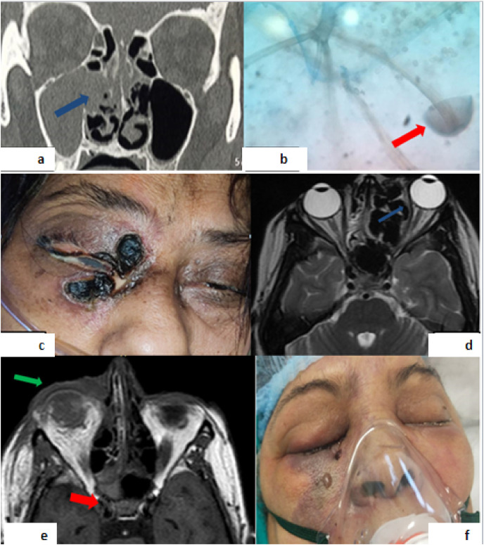
Coronavirus disease 2019 (COVID-19), may present with a myriad of clinical manifestations and complications. Patients with COVID-19 are at increased risk of pulmonary thromboembolism, acute cardiac injury, arrhythmias, acute stroke, and secondary infections. Mucormycosis is a catastrophic fungal infection characterized by vascular invasion, thrombosis, and necrosis of tissues. We report five cases of COVID-19 infection, who developed rhino-orbital mucormycosis, during the course of treatment. Early recognition of this life-threatening infection is the key to allow for optimal treatment and improved outcomes.

Keywords: COVID-19; Mucormycosis; Rajasthan.


Figures

Fig. 1
Fig. 1
Fig. 1a. CT scan showing mucoperiosteal thickening in bilateral maxillary, ethmoid, left frontal sinuses, and blocked right osteomeatal opening (blue arrow). Fig. 1b. Lactophenol cotton blue mount of nasal discharge showing broad aseptate hyphae with sporangiospore (red arrow) of rhizopus. Fig. 1c. Image of Case 3 showing right periorbital swelling, black patch on right lids. Fig. 1d. T2W MRI showing hypointense lesion causing expansion of nasal cavity, erosion of ethmoid sinus and medial wall of left orbit (blue arrow) causing mass effect on left medial rectus muscle and proptosis of left eye ball. Fig. 1e. Mild proptosis (green arrow), pre-orbital soft tissue swelling and altered shape of right orbit, thickening and peri-neural fat stranding of right optic nerve, and bulky right cavernous sinus (red arrow) suggestive of right pre and post septal orbital cellulitis, endo-ophthalmitis, and right cavernous thrombosis. Fig. 1f. Image of Case 5 showing bilateral facial and eye (right > left) swelling with necrosis of surrounding skin.

References

    1. Werthman-ehrenreich A. Mucormycosis with orbital compartment syndrome in a patient with COVID-19. Am J Emerg Med. 2020 doi: 10.1016/j.ajem.2020.09.032. - DOI - PMC - PubMed
    1. Song G., Liang G., Liu W. Fungal Co-infections associated with global COVID-19 Pandemic : a clinical and diagnostic perspective from China. Mycopathologia. 2020;185(4):599–606. - PMC - PubMed
    1. Farmakiotis D., Kontoyiannis D.P. Mucormycoses. Infect Dis Clin North Am. 2016;30:143–163. - PubMed
    1. Balai E., Mummadi S., Jolly K., Darr A., Aldeerawi H. Rhinocerebral mucormycosis: a ten-year single centre case series. Cureus. 2020;12(11) - PMC - PubMed
    1. Chen N., Zhou M., Dong X., Qu J., Gong F., Han Y., et al. Epidemiological and clinical characteristics of 99 cases of 2019 novel coronavirus pneumonia in Wuhan, China: a descriptive study. Lancet. 2020;395:507–513. - PMC - PubMed

Publication types