. 2021 Oct;75(4):888-899.
doi: 10.1016/j.jhep.2021.05.018. Epub 2021 May 28.

Focal adhesion kinase (FAK) promotes cholangiocarcinoma development and progression via YAP activation

Affiliations

Focal adhesion kinase (FAK) promotes cholangiocarcinoma development and progression via YAP activation

Xinhua Song et al. J Hepatol. 2021 Oct.

Abstract

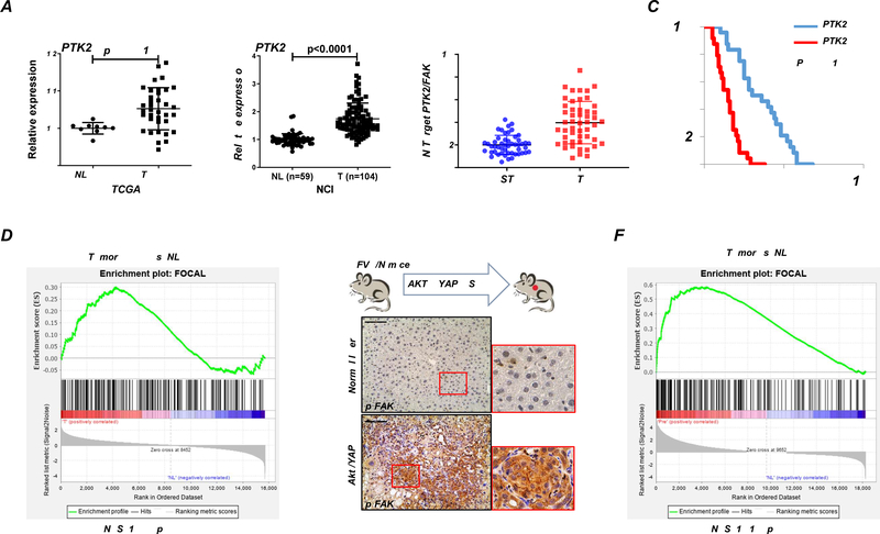
Background & aims: Focal adhesion kinase (FAK) is a non-receptor tyrosine kinase that is upregulated in many tumor types and is a promising target for cancer therapy. Herein, we elucidated the functional role of FAK in intrahepatic cholangiocarcinoma (iCCA) development and progression.

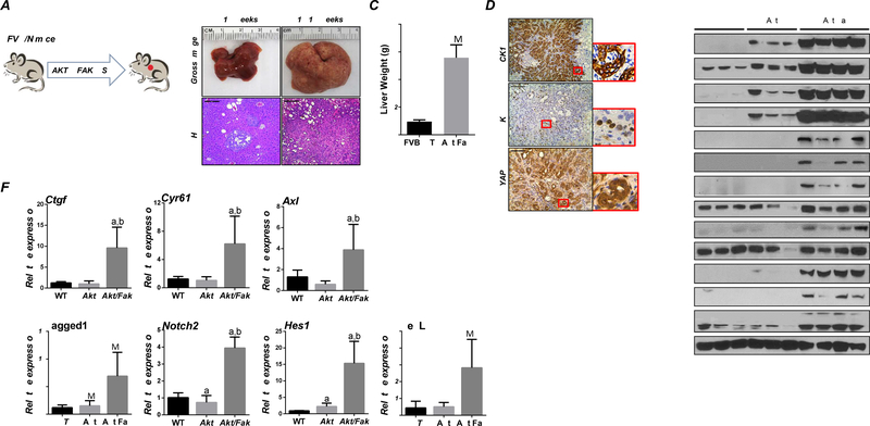
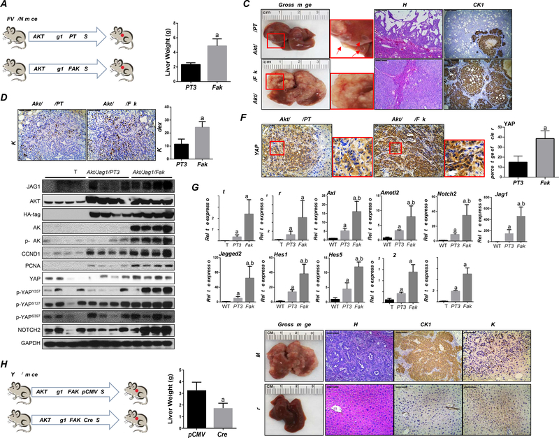
Methods: Expression levels and activation status of FAK were determined in human iCCA samples. The functional contribution of FAK to Akt/YAP murine iCCA initiation and progression was investigated using conditional Fak knockout mice and constitutive Cre or inducible Cre mice, respectively. The oncogenic potential of FAK was further examined via overexpression of FAK in mice. In vitro cell line studies and in vivo drug treatment were applied to address the therapeutic potential of targeting FAK for iCCA treatment.

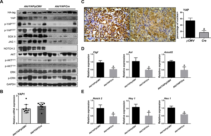
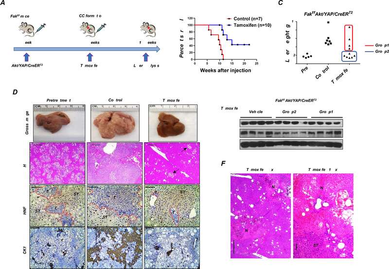
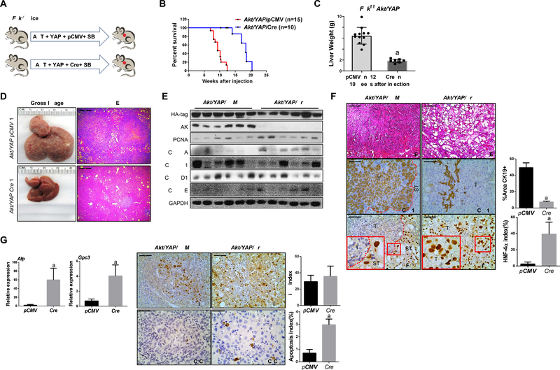
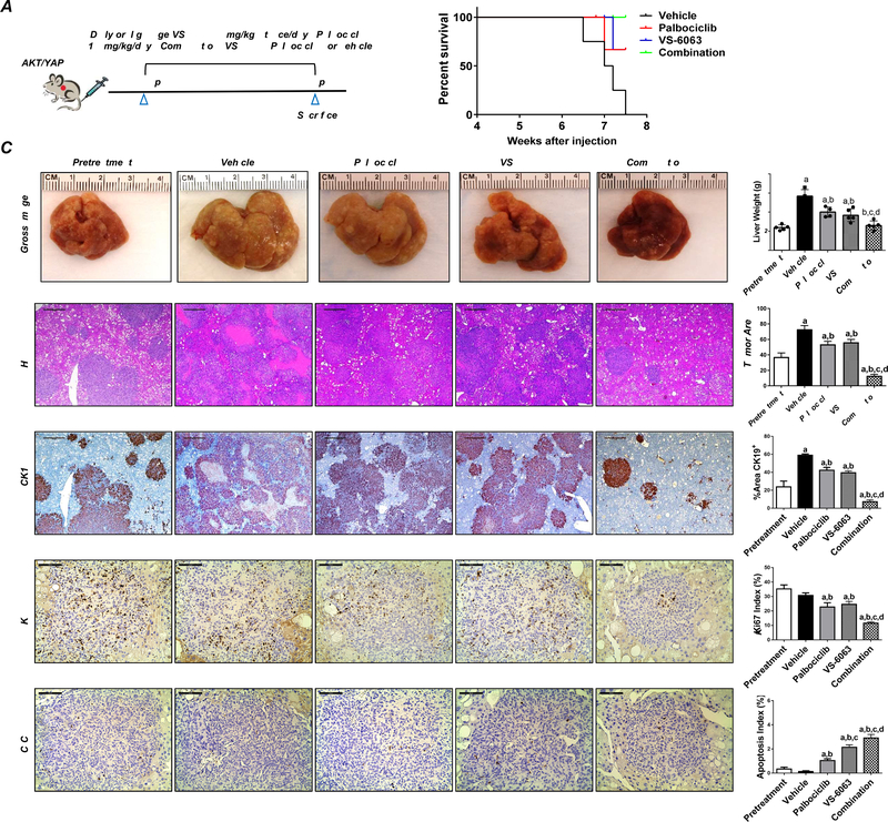
Results: FAK was ubiquitously upregulated and activated in iCCA lesions. Ablation of FAK strongly delayed Akt/YAP-driven mouse iCCA initiation. FAK overexpression synergized with activated AKT to promote iCCA development and accelerated Akt/Jag1-driven cholangiocarcinogenesis. Mechanistically, FAK was required for YAP(Y357) phosphorylation, supporting the role of FAK as a central YAP regulator in iCCA. Significantly, ablation of FAK after Akt/YAP-dependent iCCA formation strongly suppressed tumor progression in mice. Furthermore, a remarkable iCCA growth reduction was achieved when a FAK inhibitor and palbociclib, a CDK4/6 inhibitor, were administered simultaneously in human iCCA cell lines and Akt/YAP mice.

Conclusions: FAK activation contributes to the initiation and progression of iCCA by inducing the YAP proto-oncogene. Targeting FAK, either alone or in combination with anti-CDK4/6 inhibitors, may be an effective strategy for iCCA treatment.

Lay summary: We found that the protein FAK (focal adhesion kinase) is upregulated and activated in human and mouse intrahepatic cholangiocarcinoma samples. FAK promotes intrahepatic cholangiocarcinoma development, whereas deletion of FAK strongly suppresses its initiation and progression. Combined FAK and CDK4/6 inhibitor treatment had a strong anti-cancer effect in in vitro and in vivo models. This combination therapy might represent a valuable and novel treatment against human intrahepatic cholangiocarcinoma.

Keywords: FAK; YAP; cancer; intrahepatic cholangiocarcinoma; targeted therapy.


Conflict of interest statement

Conflict of interest The authors have no conflict of interest to disclose. Please refer to the accompanying ICMJE disclosure forms for further details.

Figures

Figure 1.
Figure 1.. FAK is activated in human iCCA patients and Akt/YAP-induced iCCA mouse lesions.
(A) PTK2 mRNA levels in human intrahepatic cholangiocarcinoma (iCCA) from The Cancer Genome Atlas (TCGA) and National Cancer Institute (NCI) datasets. (B) Quantitative real-time RT-PCR analysis of PTK2 expression in iCCA (n = 50) and corresponding non-tumorous surrounding liver tissues (ST; n = 50) from our cohort. (C) Kaplan–Meier survival curves of human iCCA specimens from our cohort with high and low PTK2 mRNA levels, showing the unfavorable outcome of patients with elevated expression of this gene. (D) GSEA analysis of FAK pathway-related proteins in iCCA from TCGA. (E-F) FAK activation in the Akt/YAP-induced iCCA mouse model.
Figure 2.
Figure 2.. Deletion of FAK suppresses iCCA development in Akt/YAP mice.
(A) Study design. (B) Survival analysis of Fakf/f mice bearing Akt/YAP/pCMV (n =15) and Akt/YAP/Cre (n = 10) tumors. (C) Liver weight of Akt/YAP/pCMV and Akt/YAP/Cre mice at 10 weeks post hydrodynamic injection. (D) Gross image and H&E staining of Akt/YAP/pCMV and Akt/YAP/Cre mouse livers. (E) Western blot analysis of iCCA tissues from Akt/YAP-pCMV and Cre Fakf/f mice. GAPDH was the loading control. (F) H&E and IHC staining of CK19 and HNF-4α in Akt/YAP/pCMV and Akt/YAP/Cre Fakf/f mice, respectively. CK19 staining was quantified as the percentage of the positive staining area of the whole tumor section area. HNF-4α positive cells were quantified as HNF-4α index. (G) Relative mRNA expressions of AFP and GPC3 were analyzed using the −ΔΔCt method and presented as mean ± SD. (H) Immunohistochemistry of Ki-67 and C-C-3 was performed in Akt/YAP/pCMV and Akt/YAP/Cre Fakf/f mice. Ki-67 and C-C-3 positive cells were quantified using the Image J. Tukey–Kramer test: at least P < 0.005; a, vs. Akt/YAP/pCMV; b, vs. Akt/YAP/Cre. Abbreviations: C-C-3, Cleaved Caspase 3; T, tumor; ST, surrounding tissue. Scale bar: 100 μm for 200×.
Figure 3.
Figure 3.. YAP signaling inactivation in iCCA from FAK conditional knockout Akt/YAP mice.
(A) Western blot analysis of lysates from Akt/YAP/pCMV and Akt/YAP/Cre Fakf/f mice. (B) Levels of human Yap1 mRNA in Akt/YAP/pCMV and Akt/YAP/Cre mice. (C) Knockout of FAK reduced YAP nuclear translocation as shown by immunohistochemistry and the percentage of positive nuclear staining. (D) YAP and (E) NOTCH signaling inactivation at the transcriptional level, as assessed by qPCR. Tukey–Kramer test: at least P < 0.05; a, vs Akt/YAP-pCMV; b, vs Akt/YAP-Cre.
Figure 4.
Figure 4.. Co-expression of FAK and AKT leads to iCCA formation in mice.
(A) Study design. (B) Gross image and H&E staining of Akt/Fak mouse livers. (C) Liver weight of Akt/Fak mice at 18–19 weeks post-injection. (D) Immunohistochemistry of CK19, Ki-67, and YAP in Akt/Fak mice. (E) Western blot analysis of FVB mice and Akt/Fak mouse model liver tissues. (F) YAP and NOTCH signaling activation at the transcriptional level, as assessed by qPCR. Tukey–Kramer test: at least P < 0.05; a, vs FVB WT; b, vs Akt.
Figure 5.
Figure 5.. FAK accelerates tumor development in Akt/Jag 1 mice.
(A) Study design. (B) Akt/Jag1/pT3 and Akt/Jag1/Fak mice’s liver weight at ~10 weeks post hydrodynamic injection. (C) Gross image, H&E, CK19, and (D) Ki-67 staining in Akt/Jag1/PT3 and Akt/Jag1/Fak mice. Ki-67 positive cells were quantified using Image J software. (E) Representative Western Blot analysis of relative pathways in Akt/Jag1/PT3 and Akt/Jag1/Fak mice. (F) Immunohistochemistry of YAP in Akt/Jag1/PT3 and Akt/Jag1/Fak mice. (G) YAP and NOTCH signaling activation at the transcriptional level was assessed by qPCR. (H) Study design in Yapf/f mice. (I-J) Liver weight (I) and gross image, H&E, CK19, and Ki-67 staining (J) of Akt/Jag1/Fak-pCMV and -Cre Yapf/f mice ~9 weeks post-injection. Tukey–Kramer test: at least P < 0.005; (G) a, vs WT; b, vs Akt/Jag1/Fak/pCMV, (I) a, vs Akt/Jag1/Fak/pCMV.
Figure 6.
Figure 6.. FAK downregulation after tumor formation using the CK19-CreERT2 Tamoxifen system inhibits tumor progression.
(A) Study design. (B) Survival analysis of Fakf/f mice bearing Akt/YAP/CreERT2 tumors treated with Tamoxifen or vehicle (Control). (C) Liver weight, (D) Gross image, H&E staining, HNF-4α, and CK19 immunohistochemistry of livers from pretreatment, control, and Tamoxifen groups. Arrows indicate tiny lesions consisting of small cell clusters, which were typically conserved in Tamoxifen-treated mice. (E) FAK Western blot analysis was performed in Fakf/f mice bearing Akt/YAP/CreERT2 tumors sensitized with Tamoxifen (Group2 in C) or not (Group1 in C). (F) Large areas of necrosis (N) in livers from Group2 mice, as shown at two magnifications. Abbreviations: T, tumor; ST, surrounding tissue.
Figure 7.
Figure 7.. Combining FAK inhibitor VS-6063 and Palbociclib has potent anti-neoplastic activity in iCCA lesions from Akt/YAP mice.
(A) Study design. (B) Survival curve, (C) Gross images and H&E, Ki-67, CK19, and C-C-3 staining of livers from pretreatment, vehicle-, VS-6063-, Palbociclib-, and VS-6063/Palbociclib-treated Akt/YAP mice. Asterisks indicate necrotic areas. H&E, CK19: Magnification ×40; scale bar =500μm. Ki-67, C-C-3: Magnification ×200; scale bar =100μm. CK19 positive staining, tumor area on H&E-stained slides, Ki-67, and C-C-3–positive cells were counted and quantified by Image J software. Tukey–Kramer test: at least P < 0.05. a, vs. Pretreatment; b, vs. Vehicle; c, vs. Palbociclib; d, vs. VS-6063. Abbreviations: C-C-3, cleaved caspase 3.

Comment in

References

    1. Rizvi S, Khan SA, Hallemeier CL, Kelley RK, Gores GJ. Cholangiocarcinoma - evolving concepts and therapeutic strategies. Nature reviews Clinical oncology 2018;15:95–111. - PMC - PubMed
    1. Sulzmaier FJ, Jean C, Schlaepfer DD. FAK in cancer: mechanistic findings and clinical applications. Nature reviews Cancer 2014;14:598–610. - PMC - PubMed
    1. Panera N, Crudele A, Romito I, Gnani D, Alisi A. Focal Adhesion Kinase: Insight into Molecular Roles and Functions in Hepatocellular Carcinoma. International journal of molecular sciences 2017;18;99. - PMC - PubMed
    1. Lee BY, Timpson P, Horvath LG, Daly RJ. FAK signaling in human cancer as a target for therapeutics. Pharmacology & therapeutics 2015;146:132–149. - PubMed
    1. Mohanty A, Pharaon RR, Nam A, Salgia S, Kulkarni P, Massarelli E. FAK-targeted and combination therapies for the treatment of cancer: an overview of phase I and II clinical trials. Expert opinion on investigational drugs 2020;29:399–409. - PubMed

Publication types

MeSH terms

Substances