. 2021 Sep 15:1767:147539.
doi: 10.1016/j.brainres.2021.147539. Epub 2021 May 28.

Responses of neurons in rostral ventromedial medulla to nociceptive stimulation of craniofacial region and tail in rats

Affiliations

Responses of neurons in rostral ventromedial medulla to nociceptive stimulation of craniofacial region and tail in rats

Jing-Shi Tang et al. Brain Res. .

Abstract

The rostral ventromedial medulla (RVM) plays a key role in the endogenous modulation of nociceptive transmission in the central nervous system (CNS). The primary aim of this study was to examine whether the activities of RVM neurons were related to craniofacial nociceptive behaviour (jaw-motor response, JMR) as well as the tail-flick response (TF). The activities of RVM neurons and TF and JMR evoked by noxious heating of the tail or perioral skin were recorded simultaneously in lightly anaesthetized rats. Tail or perioral heating evoked the TF and JMR, and the latency of the JMR was significantly shorter (P < 0.001) than that of the TF. Of 89 neurons recorded in RVM, 40 were classified as ON-cells, 27 as OFF-cells, and 22 as NEUTRAL-cells based on their responsiveness to heating of the tail. Heating at either site caused an increase in ON-cell and decrease in OFF-cell activity before the occurrence of the TF and JMR, but did not alter the activity of NEUTRAL cells. Likewise, noxious stimulation of the temporomandibular joint had similar effects on RVM neurons. These findings reveal that the JMR is a measure of the excitability of trigeminal and spinal nociceptive circuits in the CNS, and that the JMR as well as TF can be used for studying processes related to descending modulation of pain. The findings also support the view that RVM ON- and OFF-cells play an important role in the elaboration of diverse nociceptive behaviours evoked by noxious stimulation of widely separated regions of the body.

Keywords: Craniofacial; Descending modulation; Neurons; Pain; Rostral ventromedial medulla.


Conflict of interest statement

None of the authors has a conflict of interest to declare.

Figures
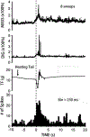
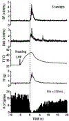
Fig. 1.
Fig. 1.
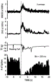
An ON-cell excited by noxious thermal stimulation of the tail. The neuronal ongoing activity was increased prior to the occurrence of the tail-flick response (TF, force scaled in g) and jaw-motor response (JMR; represented by anterior digastric [DIG] and masseter [MASS] muscle EMG responses). The force of the tail flick movement was monitored during heating of the tail produced by immersing the distal portion of the tail into a hot water bath at 55°C. The changes in neuronal activity were judged as a meaningful increase only if the firing rate increased to 150% or more of the baseline value that was an average of the baseline firing rate during the 3–5 min period before the sequence of heating stimuli. Responses to eight consecutive heating stimuli are superimposed. The dotted line indicates the onset of the TF.
Fig. 2.
Fig. 2.
An OFF-cell inhibited by noxious thermal stimulation of the tail. The neuronal ongoing activity abruptly stopped prior to the occurrence of the tail-flick response (TF, force scaled in g) and jaw-motor response (JMR; represented by anterior digastric [DIG] and masseter [MASS] muscle EMG responses). The force of the tail flick movement was monitored during heating of the tail by immersing the distal portion of the animal’s tail into a hot water bath at 55°. The changes in neuronal activity were judged as a meaningful decrease only if the firing rate decreased below 50% of the baseline value that was an average of the baseline firing rate during the 3-5 min period before heating. Responses to three consecutive heating stimuli are superimposed. The dotted line indicates the onset of the TF.
Fig. 3.
Fig. 3.
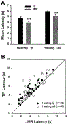
A, Comparison between mean latency of tail-flick response (TF) and mean latency of jaw-motor response (JMR). The TF (black bar) and JMR (shaded bar) were evoked during alternate noxious heating of the tail and perioral skin (upper lip). The data are presented as mean±SEM. The difference in TF and JMR latencies is statistically significant (***P<0.001); B, Individual JMR latency values plotted against their TF latency values, either during heating of the lip (●) or the tail (○). Note that in both heating conditions, most points are located to the left of the diagonal line.
Fig. 4.
Fig. 4.
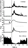
An ON-cell excited by noxious thermal stimulation of the perioral skin (upper lip). The neuronal ongoing activity was increased prior to the occurrence of the tail-flick response (TF, force scaled in g) and jaw-motor response (JMR; represented by anterior digastric [DIG] and masseter [MASS] muscle EMG responses). The TF and ON-cell activity were assessed as in Fig. 1. Responses to eight consecutive heating stimuli are superimposed. The dotted line indicates the onset of the JMR.
Fig. 5.
Fig. 5.
An OFF-cell inhibited by noxious thermal stimulation of the perioral skin (upper lip). The neuronal ongoing activity abruptly stopped prior to the occurrence of the tail-flick response (TF, force scaled in g) and jaw-motor response (JMR; represented by anterior digastric [DIG] and masseter [MASS] muscle EMG responses). The TF and OFF-cell activity were assessed as in Fig. 2. Responses to three consecutive stimuli are superimposed. The dotted line indicates the onset of the JMR.
Fig. 6.
Fig. 6.
An OFF-cell inhibited by noxious thermal stimulation of the left hindpaw (LHP). The neuronal ongoing activity abruptly stopped prior to the occurrence of the tail-flick response (TF), hindpaw withdrawal response (HWR) as well as jaw-motor response (JMR). The HWR is represented by biceps femoris (BF) muscle EMG responses, and the JMR is represented by anterior digastric (DIG) muscle EMG responses). The TF and OFF-cell activity were assessed as in Fig. 2. Responses to three consecutive heating stimuli are superimposed. Note that there were no clear differences in the time intervals between the changes of neuronal activity and the onsets of the HWR, JMR and TF. The dotted line indicates the onset of the HWR.
Fig. 7.
Fig. 7.
Reconstruction of the location of each single unit recording site (for the group of 19 rats) in the rostral ventromedial medulla (RVM). The recording sites are shown at two anterior-posterior levels. The sites of neurons classified as ON-cells, OFF-cells and NEUTRAL-cells according to their response to noxious thermal stimulation applied to the tail are indicated by the symbols shown. The number indicated at the lower right part of each diagram refers to the anterior-posterior level in relation to the bregma (Paxinos and Watson, 1998). Abbreviations: 7, Facial nucleus; 7n, facial nerve root; Sp5, trigeminal spinal tract nucleus.
Fig. 8.
Fig. 8.
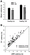
Bar graphs comparing the time intervals between the onsets of inhibition of OFF-cells and excitation of ON-cells and the tail-flick response (TF) or the jaw-motor response (JMR). The TF (black bar) and JMR (shaded bar) were evoked by heating of either the tail (A) or the perioral skin (upper lip; B). The time intervals between the onset of changes of neuronal activity and the onset of the JMR were significantly shorter than those for the TF. Data are presented as mean±SEM (**P<0.01; *** P<0.001).

Similar articles

Cited by

References

    1. Bannon AW, Malmberg AB, 2007. Models of nociception: hot-plate, tail-flick, and formalin tests in rodents. Curr. Protoc. Neurosci. Chapter 8, Unit 8.9 doi:10.1002/0471142301.ns0809s41. - DOI - PubMed
    1. Basbaum AI, Clanton CH, Fields HL, 1976. Opiate and stimulus-produced analgesia: functional anatomy of a medullospinal pathway. Proc. Natl. Acad. Sci. U. S. A 73(12),4685–4688. doi:10.1073/pnas.73.12.4685. - DOI - PMC - PubMed
    1. Basbaum AI, Clanton CH, Fields HL, 1978. Three bulbospinal pathways from the rostral medulla of the cat: an autoradiographic study of pain modulating systems. J. Comp. Neurol 178(2), 209–224. doi:10.1002/cne.901780203. - DOI - PubMed
    1. Berge OG, Garcia-Cabrera I, Hole K, 1988. Response latencies in the tail-flick test depend on tail skin temperature. Neurosci. Lett 86(3),284–288. doi:10.1016/0304-3940(88)90497-1. - DOI - PubMed
    1. Bouhassira D, Chitour D, Villanueva L, Le Bars D, 1993. Morphine and diffuse noxious inhibitory controls in the rat: effects of lesions of the rostral ventromedial medulla. Eur. J. Pharmacol 232(2–3),207–215. doi:10.1016/0014-2999(93)90775-d. - DOI - PubMed

Publication types