

Long-Term Observation and Sequencing Analysis of SKPs-Derived Corneal Endothelial Cell-Like Cells for Treating Corneal Endothelial Dysfunction
- PMID: 34053246
- PMCID: PMC8182626
- DOI: 10.1177/09636897211017830
Long-Term Observation and Sequencing Analysis of SKPs-Derived Corneal Endothelial Cell-Like Cells for Treating Corneal Endothelial Dysfunction
Abstract
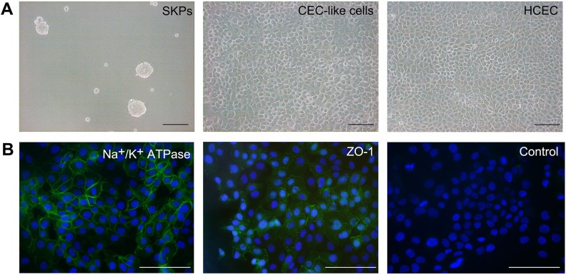
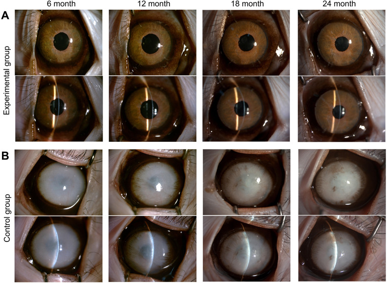
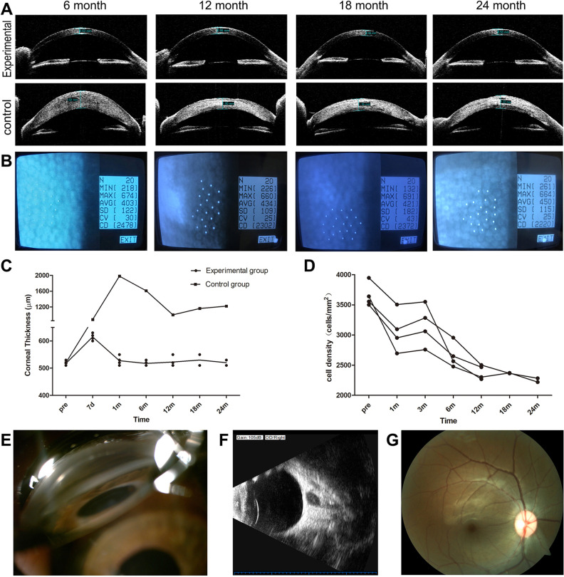
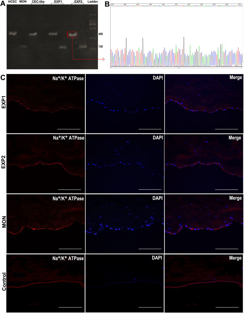
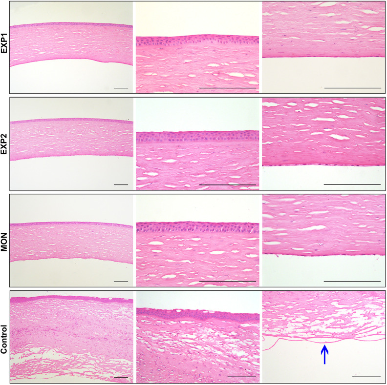
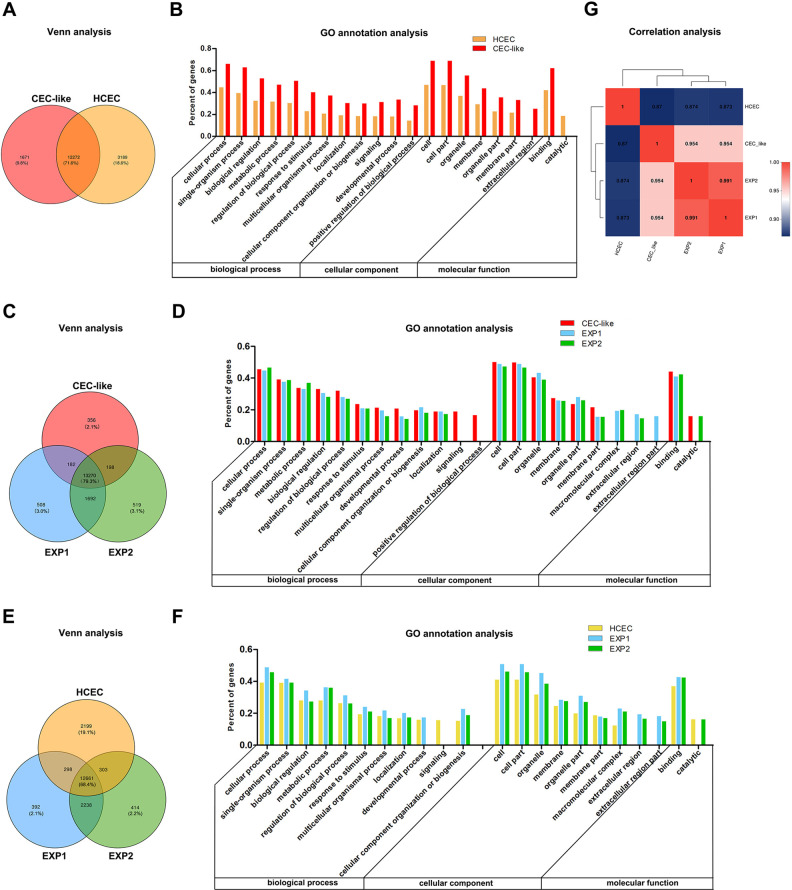
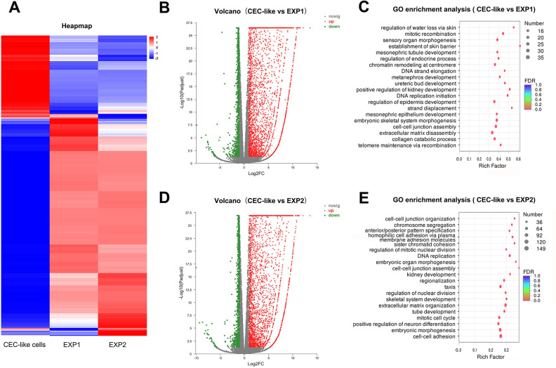
Corneal endothelial dysfunction is a principal cause of visual deficiency. Corneal transplantation is the most effective treatment for corneal endothelial dysfunction. However, a severe shortage of available donor corneas or human corneal endothelial cells (HCECs) remains a global challenge. Previously, we acquired corneal endothelial cell-like cells (CEC-like cells) derived from human skin-derived precursors (SKPs). CEC-like cells were injected into rabbit and monkey corneal endothelial dysfunction models and exerted excellent therapeutic effect. In this study, we prolonged the clinical observation in the monkey experiment for 2 years. Polymerase chain reaction (PCR) and DNA sequencing were carried out to confirm the existence of CEC-like cells. Histological examinations were carried out to show the corneal morphology. Further transcriptome sequencing was also carried out on HCEC, CEC-like cells before transplantation and after transplantation. We found that the monkeys cornea remained transparent and normal thickness. The total endothelial cell density decreased gradually, but tended to be stable and remained in a normal range during 2-year observation. The CEC-like cells persist during observation and could adapt to the microenvironment after transplantation. The gene expression pattern of CEC-like cells was similar to HCEC and changed slightly after transplantation. In conclusion, this study presented a brand-new insight into CEC-like cells and further provided a promising prospect of cell-based therapy for corneal endothelial dysfunction. The renewable cell source, novel derivation method and simple treatment strategy may be clinically applied in regenerative medicine in the future.
Keywords: cell-based therapy; corneal endothelial cell-like cells; corneal endothelial dysfunction; skin-derived precursors; transcriptome sequencing.
Conflict of interest statement
Figures
Similar articles
-
Construction of tissue engineered cornea with skin-derived corneal endothelial-like cell and mechanism research for the cell differentiation.Front Med (Lausanne). 2024 Sep 2;11:1448248. doi: 10.3389/fmed.2024.1448248. eCollection 2024. Front Med (Lausanne). 2024. PMID: 39286645 Free PMC article.
-
Long-term observation after transplantation of cultured human corneal endothelial cells for corneal endothelial dysfunction.Stem Cell Res Ther. 2022 Jun 3;13(1):228. doi: 10.1186/s13287-022-02889-x. Stem Cell Res Ther. 2022. PMID: 35659288 Free PMC article.
-
Therapy of corneal endothelial dysfunction with corneal endothelial cell-like cells derived from skin-derived precursors.Sci Rep. 2017 Oct 17;7(1):13400. doi: 10.1038/s41598-017-13787-1. Sci Rep. 2017. PMID: 29042661 Free PMC article.
-
[Cultured human corneal endothelial cell transplantation].Nippon Ganka Gakkai Zasshi. 2006 Nov;110(11):879-97. Nippon Ganka Gakkai Zasshi. 2006. PMID: 17134036 Review. Japanese.
-
[Transplantation of corneal endothelial cells].Nippon Ganka Gakkai Zasshi. 2002 Dec;106(12):805-35; discussion 836. Nippon Ganka Gakkai Zasshi. 2002. PMID: 12610838 Review. Japanese.
Cited by
-
Dermal Papilla Cells: From Basic Research to Translational Applications.Biology (Basel). 2024 Oct 20;13(10):842. doi: 10.3390/biology13100842. Biology (Basel). 2024. PMID: 39452150 Free PMC article. Review.
-
Long-term corneal recovery by simultaneous delivery of hPSC-derived corneal endothelial precursors and nicotinamide.J Clin Invest. 2022 Jan 4;132(1):e146658. doi: 10.1172/JCI146658. J Clin Invest. 2022. PMID: 34981789 Free PMC article.
-
Construction of tissue engineered cornea with skin-derived corneal endothelial-like cell and mechanism research for the cell differentiation.Front Med (Lausanne). 2024 Sep 2;11:1448248. doi: 10.3389/fmed.2024.1448248. eCollection 2024. Front Med (Lausanne). 2024. PMID: 39286645 Free PMC article.
-
Research on the construction of corneal endothelium transplantation with acellular amniotic membrane as a scaffold.Front Med (Lausanne). 2025 Jul 1;12:1592123. doi: 10.3389/fmed.2025.1592123. eCollection 2025. Front Med (Lausanne). 2025. PMID: 40665979 Free PMC article.
-
Biomedical Application of MSCs in Corneal Regeneration and Repair.Int J Mol Sci. 2025 Jan 15;26(2):695. doi: 10.3390/ijms26020695. Int J Mol Sci. 2025. PMID: 39859409 Free PMC article. Review.
References
-
- Flaxman SR, Bourne RRA, Resnikoff S, Ackland P, Braithwaite T, Cicinelli MV, Das A, Jonas JB, Keeffe J, Kempen JH, Leasher J, Leasher J, Limburg H, Naidoo K, Pesudovs K, Silvester A, Stevens GA, Tahhan N, Wong TY, Taylor HR, Vision Loss Expert Group of the Global Burden of Disease S. Global causes of blindness and distance vision impairment 1990-2020: a systematic review and meta-analysis. Lancet Glob Health. 2017;5(12):e1221–e1234. - PubMed
-
- Lee JG, Ko MK, Kay EP. Endothelial mesenchymal transformation mediated by IL-1beta-induced FGF-2 in corneal endothelial cells. Exp Eye Res. 2012;95(1):35–39. - PubMed
-
- Rolev K, Coussons P, King L, Rajan M. Experimental models of corneal endothelial cell therapy and translational challenges to clinical practice. Exp Eye Res. 2019;188:107794. - PubMed
-
- Ono T, Ishiyama S, Hayashidera T, Mori Y, Nejima R, Miyata K, Amano S. Twelve-year follow-up of penetrating keratoplasty. Jpn J Ophthalmol. 2017;61(2):131–136. - PubMed
-
- Fuest M, Ang M, Htoon HM, Tan D, Mehta JS. Long-term visual outcomes comparing descemet stripping automated endothelial keratoplasty and penetrating keratoplasty. Am J Ophthalmol. 2017;182:62–71. - PubMed
Publication types
MeSH terms
LinkOut - more resources
Full Text Sources
Other Literature Sources
