. 2021 Dec;12(1):1452-1468.
doi: 10.1080/21505594.2021.1929037.

Reduction of endotoxicity in Bordetella bronchiseptica by lipid A engineering: Characterization of lpxL1 and pagP mutants

Affiliations

Reduction of endotoxicity in Bordetella bronchiseptica by lipid A engineering: Characterization of lpxL1 and pagP mutants

Jesús Pérez-Ortega et al. Virulence. 2021 Dec.

Abstract

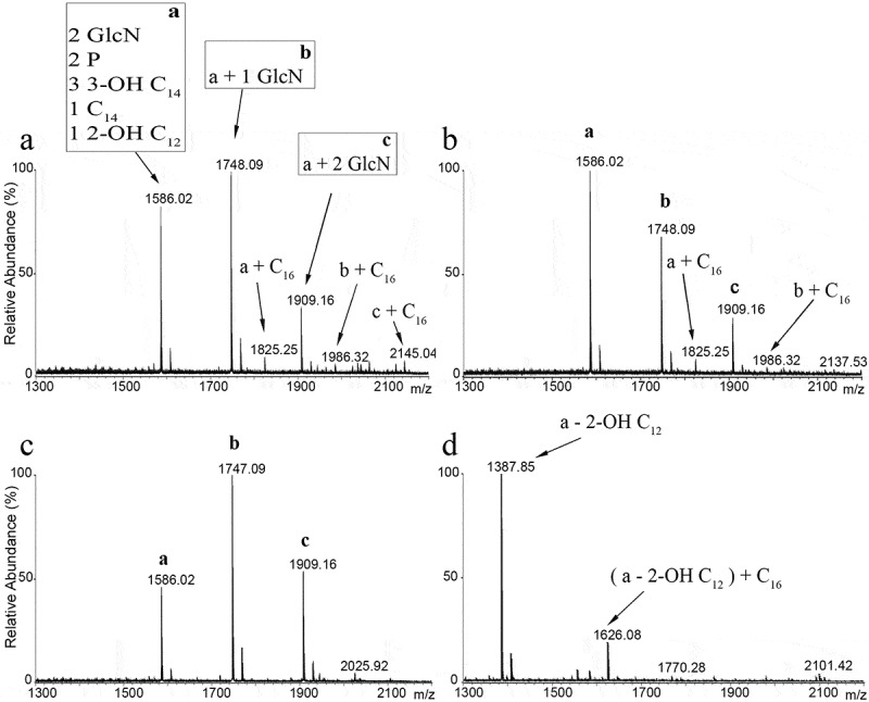
Whole-cell vaccines against Gram-negative bacteria commonly display high reactogenicity caused by the endotoxic activity of lipopolysaccharide (LPS), one of the major components of the bacterial outer membrane. Underacylation of the lipid A moiety of LPS has been related with reduced endotoxicity in several Gram-negative species. Here, we evaluated whether the inactivation of two genes encoding lipid A acylases of Bordetella bronchiseptica, i.e. pagP and lpxL1, could be used for the development of less reactogenic vaccines against this pathogen for livestock and companion animals. Inactivation of pagP resulted in the loss of the secondary palmitate chain at position 3' of lipid A, but hardly affected the potency of the LPS to activate the Toll-like receptor 4 (TLR4). Inactivation of lpxL1 resulted in the loss of the secondary 2-hydroxy laurate group present at position 2 of lipid A and, unexpectedly, in the additional loss of the glucosamines that decorate the phosphate groups at positions 1 and 4' and in an increase in LPS molecules carrying O-antigen. The resulting LPS showed greatly reduced potency to activate TLR4 in HEK-Blue reporter cells expressing human or mouse TLR4 as well as in porcine macrophages. Characterization of the lpxL1 mutant revealed many pleiotropic phenotypes, including increased resistance to SDS and rifampicin, increased susceptibility to cationic antimicrobial peptides, decreased auto-aggregation and biofilm formation, and a tendency to decreased infectivity of macrophages, which are all related to the altered LPS structure. We suggest that the lpxL1 mutant will be useful for the generation of safer vaccines.

Keywords: Bordetella; LPS; LpxL1; PagP; TLR4; antimicrobial susceptibility; autoaggregation; biofilms; endotoxicity; vaccine.


Figures

Figure 1.
Figure 1.
Representation of the lipid A structure of B. bronchiseptica. The non-stoichiometric glucosamine and C16 acyl chain modifications are denoted by dashed bond lines. The enzymes responsible for the addition of the secondary acyl chains are indicated
Figure 2.
Figure 2.
LC-MS analysis of B. bronchiseptica lipid A. Comparison of lipid A species from B. bronchiseptica strains BB-P19 (a) and BB-D09-SR (b), and the pagP (c) and lpxL1 (d) mutant derivatives, respectively. Major peaks at m/z 1586, 1748 and 1909 were interpreted as the characteristic penta-acylated bis-phosphorylated species, and the corresponding species with one and two glucosamine substituents, respectively. Predicted divergence for the other peaks is indicated
Figure 3.
Figure 3.
Analysis of LPS modifications by SDS-PAGE. Purified LPS of B. bronchiseptica strain BB-D09-SR and of the pagP and lpxL1 mutants was analyzed by SDS-PAGE and visualized by silver staining. The identity of the various bands detected at the position of lipid A plus core sugars is discussed in the text. O-antigen-containing LPS appears as diffuse smear with lower electrophoretic mobility
Figure 4.
Figure 4.
TLR4 activation by purified LPS and whole-cell preparations of strain BB-D09-SR and its pagP and lpxL1 mutant derivatives. HEK-Blue cells expressing either m-TLR4 (left panels) or h-TLR4 (right panels) were incubated for 17 h with 10-fold serial dilutions of (a) purified LPS or (b) heat-inactivated whole cells. The concentrations of the undiluted samples are indicated in ng/ml and OD600 units in panels A and B, respectively. Graphs show mean ± SEM of SEAP activity measured at OD405 from the supernatants of three independent experiments performed in duplicate. In the stimulation assays with LPS, statistical comparison showed significant differences relative to the wild type only for the lpxL1 mutant (P < 0.0001 for both m- and h-TLR4). When stimulated with whole cells, significant differences were found for the lpxL1 mutant (P < 0.0001 for both m- and h-TLR4) but also for the pagP mutant (P < 0.05 for m-TLR4 and P < 0.0001 for h-TLR4)
Figure 5.
Figure 5.
Cytokine secretion upon stimulation of PBMMs with LPS from the B. bronchiseptica strain BB-D09-SR and its pagP- and lpxL1-mutant derivatives. Secreted levels of TNF-α, IL-10, IL-8 and IL-1β by PBMMs of separate porcine individuals (n = 5) were measured after 24 h incubation with 10 ng/ml of purified LPS. Values shown are means and standard deviations from three independent experiments. Statistically significant differences compared to the wild type are indicated with asterisks (*, P < 0.05)
Figure 6.
Figure 6.
Bacterial sensitivity to SDS. Cultures of strain BB-D09-SR and its pagP- and lpxL1-mutant derivatives were incubated in duplicate with 1% SDS. After 2 h incubation, drops of 10-fold serial dilutions from these cultures were plated on BG-blood agar and incubated for 48 h. The control at the left shows the results for bacteria not exposed to SDS. A representative result of three independent experiments is shown
Figure 7.
Figure 7.
Inactivation of lpxL1 reduces settling, biofilm formation and surface hydrophobicity. (a) Macroscopic view of bacterial settling. Cultures of strain BB-D09-SR and its mutant derivatives were grown in SS medium supplemented with casamino acids and adjusted to an OD600 of 1. A photograph was taken after 5 h incubation under static conditions. (b) Absorbance was measured from the same cultures as shown in panel A. Samples were taken at 0 and 45 min, and at 2, 5 and 24 h. Graph shows mean ± SEM of absorbance relative to the t = 0 sample calculated from four independent experiments. Comparison with the parental strain showed statistically significant differences only for lpxL1 mutant (P < 0.0001). (c) Biofilm formation under static conditions. After 24 h of incubation in SS medium supplemented with casamino acids, biofilms were stained with crystal violet and quantified by measuring the OD630. Data represent means and standard deviations from three experiments performed in triplicate. Statistically significant difference compared to the wild type is indicated with asterisks (**, P < 0.01). (d) Surface hydrophobicity assessment using BATH method. Bacterial suspensions standardized at OD600 of 1 in PBS were mixed with hexadecane. Percentage hydrophobicity was calculated from OD measurements of samples from the water phase. The means and standard deviations from three experiments performed in duplicate are shown. Statistically significant difference compared to the wild type is indicated with asterisks (***, P < 0.001)
Figure 8.
Figure 8.
Bacterial survival with pro-inflammatory M1 macrophages. Suspensions of strain BB-D09-SR and its mutant derivatives were incubated with M1 macrophages at an MOI of 1 for 4 h, and CFU in different fractions were quantified. (a) Total bacterial growth (i.e. both inside and outside of the macrophages) expressed as CFU. (b) CFU in the supernatant. (c) CFU attached at the macrophage surface. (d) CFU inside macrophages. Symbols with different shapes correspond to PBMMs of separate porcine individuals (n = 7). No statistical significance was found
Figure 9.
Figure 9.
Comparison of protein content of outer-membrane preparations from B. bronchiseptica strain BB-D09-SR and its lpxL1 mutant derivative. (a) SDS-PAGE analysis of isolated outer membranes of the wild-type strain and its lpxL1 mutant derivative. Molecular weight markers are shown at the left. (b) Western blot analysis of relevant outer membrane antigens. Membranes were incubated with antibodies directed against the siderophore receptor FauA, major porin OmpP, and autotransporter BrkA

References

    1. Raetz CRH, Whitfield C.. Lipopolysaccharide endotoxins. Annu. Rev. Biochem. 2002;71(1):635–700. - PMC - PubMed
    1. Raetz CRH, Reynolds CM, Trent MS, et al. Lipid A modification systems in Gram-negative bacteria. Annu. Rev. Biochem. 2007;76(1):295–329. - PMC - PubMed
    1. Matsuura M. Structural modifications of bacterial lipopolysaccharide that facilitate Gram-negative bacteria evasion of host innate immunity. Front Immunol. 2013;4:109. - PMC - PubMed
    1. Goodnow RA. Biology of Bordetella bronchiseptica. Microbiol. Rev. 1980;44(4):722–738. - PMC - PubMed
    1. Mattoo S, Cherry JD. Molecular pathogenesis, epidemiology, and clinical manifestations of respiratory infections due to Bordetella pertussis and other Bordetella subspecies. Clin. Microbiol. Rev. 2005;18(2):326–382. - PMC - PubMed

Publication types