. 2021 May 30;27(1):53.
doi: 10.1186/s10020-021-00309-z.

Baicalin alleviates chronic obstructive pulmonary disease through regulation of HSP72-mediated JNK pathway

Affiliations

Baicalin alleviates chronic obstructive pulmonary disease through regulation of HSP72-mediated JNK pathway

Dexun Hao et al. Mol Med. .

Abstract

Background: Chronic obstructive pulmonary disease (COPD) is characterized by airway obstruction and progressive lung inflammation. As the primary ingredient of a traditional Chinese medical herb, Baicalin has been previously shown to possess anti-inflammatory abilities. Thus, the current study aimed to elucidate the mechanism by which baicalin alleviates COPD.

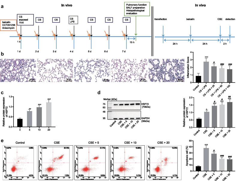
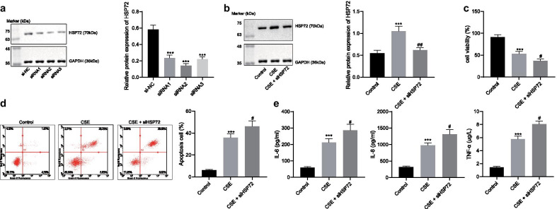
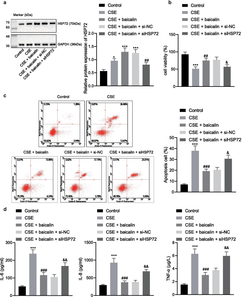
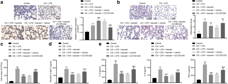
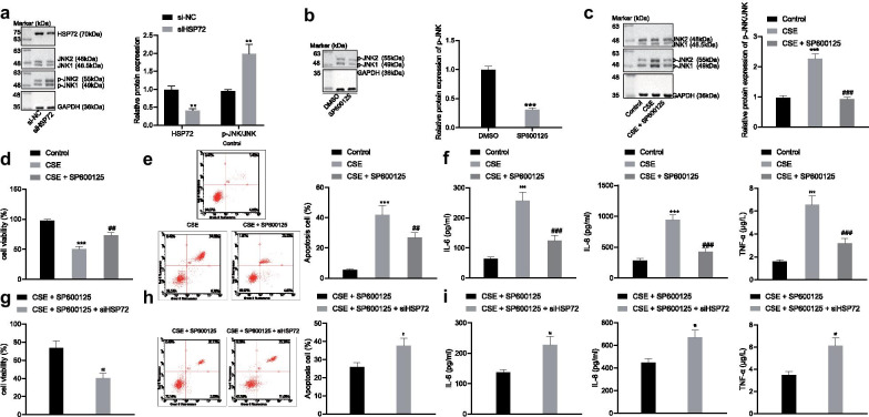
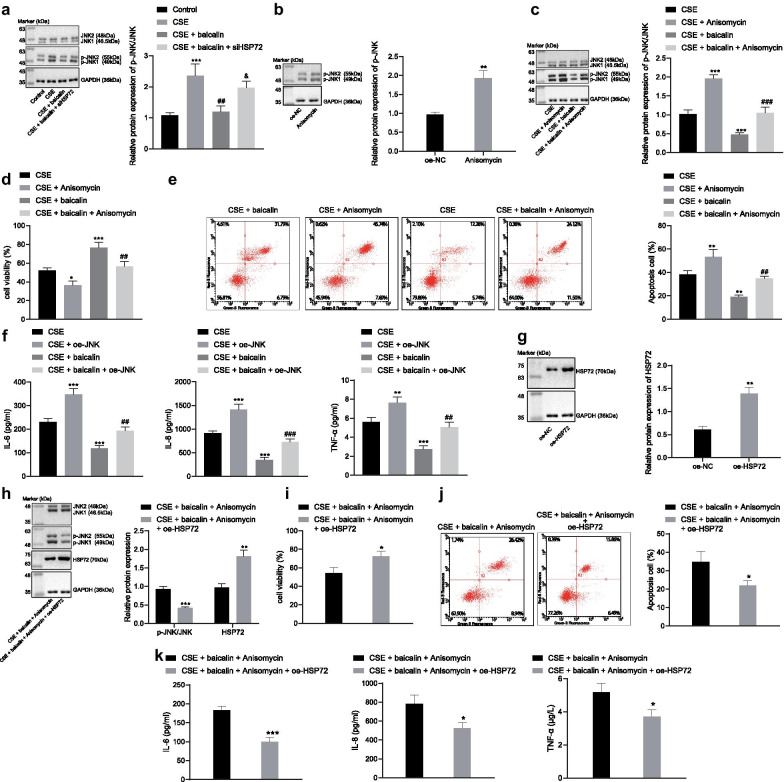
Methods: Baicalin was adopted to treat cigarette smoke in extract-exposed MLE-12 cells after which cell viability and apoptosis were determined. The production of tumor necrosis factor alpha (TNF-α), interleukin-6 (IL-6), IL-8 were determined by enzyme-linked immunoassay. A COPD mouse model was constructed via exposure to cigarette smoke and lipopolysaccharide, baicalin treatment. Lung function and inflammatory cell infiltration were determined and the production of Muc5AC, TNF-α, IL-6, IL-8 in the bronchoalveolar lavage fluid (BALF) was assayed by ELISA. The effect of HSP72 and JNK on COPD following treatment with baicalin was assessed both in vivo and in vitro by conducting loss- and gain- function experiments.

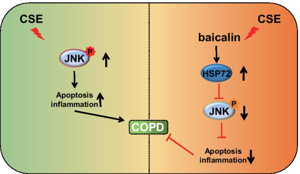
Results: Baicalin improved lung function evidenced by reduction in inflammatory cell infiltration and Muc5AC, TNF-α, IL-6 and IL-8 levels observed in BALF in mice. Baicalin was further observed to elevate cell viability while inhibited apoptosis and TNF-α, IL-6 and IL-8 levels in MLE-12 cells. Baicalin treatment increased HSP72 expression, while its depletion reversed the effect of baicalin on COPD. HSP72 inhibited the activation of JNK, while JNK activation was found to inhibit the effect of baicalin on COPD.

Conclusions: Baicalin upregulated the expression of HSP72, resulting in the inhibition of JNK signaling activation, which ultimately alleviates COPD.

Keywords: Baicalin; Chronic obstructive pulmonary disease; Heat shock protein 72; Inflammation; c-Jun N-terminal kinase signaling.


Conflict of interest statement

The authors declare that they have no competing interests.

Figures

Fig. 1
Fig. 1
Baicalin alleviates COPD and upregulates the expression of HSP72. COPD was induced by exposing the mice to CS and LPS. a Flow-process diagrams of in vivo and in vitro experiments. b Inflammatory cell infiltration in the lung was analyzed by H&E staining (×200) and the severity of inflammation was scored. c The expression of HSP72 in MLE-12 cells after baicalin treatment for 24 h, *p < 0.05 compared to mice treated with 0 µmol baicalin. d The expression of HSP72 normalized to GAPDH was analyzed using immunoblotting after MLE-12 cells were treated with baicalin for 24 h and stimulated by CSE for 2 h. e Cell apoptosis of MLE-12 cells were determined using flow cytometry. *p < 0.05, **p < 0.01, ***p < 0.0001 compared to normal mice, #p < 0.05, ##p < 0.01, ###p < 0.001 compared to mice treated with CSE. All experiments were repeated three times independently and the data were represented as mean ± standard deviation. One-way ANOVA followed by Tukey’s test was used for multiple group comparison
Fig. 2
Fig. 2
Knockdown of HSP72 exacerbates CS-induced injury on MLE-12 cells. a Relative protein expression of HSP72 following treatment of HSP72 siRNA normalized to GAPDH was evaluated by immunoblotting; *p < 0.05, **p < 0.01, ***p < 0.001 compared to cells transfected with si-NC. b MLE-12 cells was transfected with si-HSP72 and stimulated with 5% CSE, and the expression of HSP72 normalized to GAPDH was detected by immunoblotting. CSE was exposed to control and HSP72 knocked down cells. c Cell viability was tested with CCK-8 assay. d Cell apoptosis was tested with flow cytometry. e The release of IL-6, IL-8, and TNF-α was detected by ELISA, *p < 0.05, **p < 0.01, ***p < 0.001 compared to untreated cells, #p < 0.05, ##p < 0.01, ###p < 0.001 compared to cells treated with CSE. All experiments were repeated for three times independently and the data were represented as mean ± standard deviation. One-way ANOVA followed by Tukey’s test was used for multiple group comparison
Fig. 3
Fig. 3
Knockdown of HSP72 retards the effect of baicalin on the treatment of COPD. Cells were treated with CSE, CSE + baicalin, CSE + baicalin + si-NC, or CSE + baicalin + si-HSP72. Mice were treated with CS + LPS, CS + LPS + baicalin, or CS + LPS + baicalin + CCT251236. a The expression of HSP72 normalized to GAPDH was detected by immunoblotting in cells; *p < 0.05, **p < 0.01, ***p < 0.001 compared to untreated cells; #p < 0.05, ##p < 0.01, ###p < 0.001 compared to cells treated with CSE + baicalin + si-NC. b The viability of the above cells was determined by CCK-8; *p < 0.05, **p < 0.01, ***p < 0.001 compared to untreated cells; #p < 0.05, ##p < 0.01, ###p < 0.001 compared to cells treated with CSE; &p < 0.05, &&p < 0.01, &&& p < 0.001 compared to cells treated with CSE + baicalin + si-NC. c Flow cytometry was used to analyze apoptosis of the cells. d Release of inflammatory factors IL-6, IL-8 and TNF-α was measured by ELISA
Fig. 4
Fig. 4
The HSP72 inhibitor CCT251236 inhibited the therapeutic effect of baicalin on COPD in vivo. a HSP72 expression in the lung tissues of mice was tested by immunohistochemistry (×400). b Lung injury was visualized by H&E staining (×400) and lung injury was scored. c The white blood cells in the BALF were measured by ELISA. d Muc5AC expression in the BALF was determined. e Expression of IL-6, IL-8, TNF-α in the BALF was measured by ELISA. Compared with the control group, *p < 0.05, **p < 0.01, ***p < 0.001; compared with the CS + LPS group, #p < 0.05, ##p < 0.01, ###p < 0.001; compared with the CS + LPS + baicalin + vehicle group, &p < 0.05, &&p < 0.01, &&& p < 0.001. All experiments were repeated for three times independently and the data were represented as mean ± standard deviation. One-way ANOVA followed by Tukey’s test was used for multiple group comparison
Fig. 5
Fig. 5
HSP72 inhibits COPD through the inhibition of the JNK signaling pathway. a MLE-12 cells were transfected with si-HSP72 and the expression of HSP72 and JNK phosphorylation normalized to GAPDH was analyzed by immunoblotting; *p < 0.05, **p < 0.01, ***p < 0.001 compared to cells transfected with si-NC. b The extent of JNK phosphorylation normalized to GAPDH after SP600125 treatment was analyzed by immunoblotting; *p < 0.05, **p < 0.01, ***p < 0.001 compared to cells treated with DMSO. MLE-12 cells were treated with SP600125 and stimulated with CSE. c The phosphorylation of JNK normalized to GAPDH was analyzed by immunoblotting. d Cell viability was detected by CCK-8 assay. e Cell apoptosis was detected by flow cytometry. f Release of IL-6, IL-8, and TNF-α was analyzed by ELISA. *p < 0.05, **p < 0.01, ***p < 0.001 compared to untreated cells; #p < 0.05, ##p < 0.01, ###p < 0.001 compared to cells treated with CSE. MLE-12 cells were co-treated with si-HSP72 and SP600125. g Cell viability was detected by CCK-8 assay. h Cell apoptosis was detected by flow cytometry. i Release of IL-6, IL-8, and TNF-α was analyzed by ELISA. *p < 0.05, **p < 0.01,***p < 0.001 compared to CSE + SP600125. All experiments were repeated for three times independently and the data were represented as mean ± standard deviation. One-way ANOVA followed by Tukey’s test was used for multiple group comparison
Fig. 6
Fig. 6
Baicalin blocks the JNK signaling pathway and alleviates COPD in a HSP72 dependent manner. a MLE-12 cells were transfected with si-HSP72 or control vector and stimulated with CSE. The cells treated with baicalin and the phosphorylation of JNK normalized to GAPDH was analyzed by immunoblotting; *p < 0.05, **p < 0.01, ***p < 0.001 compared to untreated cells; #p < 0.05, ##p < 0.01, ###p < 0.001 compared to cells treated with CSE; &p < 0.05, ##p < 0.01, ###p < 0.001 compared to cells treated with CSE + baicalin. b The extent of JNK phosphorylation after Anisomycin treatment normalized to GAPDH was tested by immunoblotting. *p < 0.05, **p < 0.01, ***p < 0.001 compared to cells treated with vehicle. c The phosphorylation of JNK normalized to GAPDH was analyzed by immunoblotting. d Cell viability was analyzed by CCK-8. e Cell apoptosis was analyzed by flow cytometry. f Release of IL-6, IL-8 and TNF-α was analyzed ELISA. *p < 0.05, **p < 0.01, ***p < 0.001 compared to cells treated with CSE; #p < 0.05, ##p < 0.01, ###p < 0.001 compared to cells treated with CSE + baicalin. g Relative protein expression of HSP72 normalized to GAPDH after treatment of HSP72 overexpression vector was determined by immunoblotting. *p < 0.05, **p < 0.01, ***p < 0.001 compared to cells transfected with oe-NC. h Cells were co-transfected with both Anisomycin and oe-HSP72 and stimulated with CSE. The expression of HSP72 and extent of JNK phosphorylation normalized to GAPDH was analyzed by immunoblotting. i Cell viability of both JNK-activated and HSP72-overexpressed cells after treatment with baicalin was analyzed by CCK-8 assay. j Cell apoptosis of both JNK-activated and HSP72-overexpressed cells after treatment with baicalin was analyzed by flow cytometry. k Release of IL-6, IL-8 and TNF-α of both JNK-activated and HSP72-overexpressed cells after treatment with baicalin was analyzed ELISA. *p < 0.05, **p < 0.01, ***p < 0.001 compared to cells treated with CSE + baicalin + Anisomycin. All experiments were repeated for three times independently and the data were represented as mean ± standard deviation. One-way ANOVA followed by Tukey’s test was used for multiple group comparison
Fig. 7
Fig. 7
Graphic mechanism. Aqueous CSE stimulation promotes the activation of the JNK pathway thereby promoting the development of COPD, while baicalin can up-regulate the expression of HSP72, which in turn inhibits the activity of the JNK pathway and impedes the development of COPD

References

    1. Barnes PJ. Inflammatory mechanisms in patients with chronic obstructive pulmonary disease. J Allergy Clin Immunol. 2016;138(1):16–27. doi: 10.1016/j.jaci.2016.05.011. - DOI - PubMed
    1. Chen J, Zhang R, Wang J, Yu P, Liu Q, Zeng D, et al. Protective effects of baicalin on LPS-induced injury in intestinal epithelial cells and intercellular tight junctions. Can J Physiol Pharmacol. 2015;93(4):233–7. doi: 10.1139/cjpp-2014-0262. - DOI - PubMed
    1. Chen Y, Liu K, Zhang J, Hai Y, Wang P, Wang H, et al. c-Jun NH2 -Terminal Protein Kinase Phosphorylates the Nrf2-ECH Homology 6 Domain of Nuclear Factor Erythroid 2-Related Factor 2 and Downregulates Cytoprotective Genes in Acetaminophen-Induced Liver Injury in Mice. Hepatology. 2020;71(5):1787–801. doi: 10.1002/hep.31116. - DOI - PMC - PubMed
    1. Disease GBD, Injury I, Prevalence C. Global, regional, and national incidence, prevalence, and years lived with disability for 310 diseases and injuries, 1990–2015: a systematic analysis for the Global Burden of Disease Study 2015. Lancet. 2016;388(10053):1545–602. doi: 10.1016/S0140-6736(16)31678-6. - DOI - PMC - PubMed
    1. Dokladny K, Lobb R, Wharton W, Ma TY, Moseley PL. LPS-induced cytokine levels are repressed by elevated expression of HSP70 in rats: possible role of NF-kappaB. Cell Stress Chaperones. 2010;15(2):153–63. doi: 10.1007/s12192-009-0129-6. - DOI - PMC - PubMed

MeSH terms