

Ameliorative Role of Cerium Oxide Nanoparticles Against Fipronil Impact on Brain Function, Oxidative Stress, and Apoptotic Cascades in Albino Rats
- PMID: 34054412
- PMCID: PMC8163223
- DOI: 10.3389/fnins.2021.651471
Ameliorative Role of Cerium Oxide Nanoparticles Against Fipronil Impact on Brain Function, Oxidative Stress, and Apoptotic Cascades in Albino Rats
Abstract
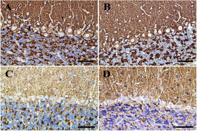
Fipronil (FIP) is an N-phenylpyrazole insecticide that is used extensively in public health and agriculture against a wide range of pests. Exposure to FIP is linked to negative health outcomes in humans and animals including promoting neuronal cell injury, which results in apoptosis through the production of reactive oxygen species (ROS). Therefore, the purpose of the current study was to investigate the neuroprotective effects of cerium oxide nanoparticles (CeNPs) on neuronal dysfunction induced by FIP in albino rats. Male rats were randomly classified into four groups: control, FIP (5 mg/kg bwt), CeNPs (35 mg/kg bwt), and FIP + CeNPs (5 (FIP) + 35 (CeNPs) mg/kg bwt), which were treated orally once daily for 28 consecutive days. Brain antioxidant parameters, histopathology, and mRNA expression of genes related to brain function were evaluated. The results revealed oxidative damage to brain tissues in FIP-treated rats indicated by the elevated levels of malondialdehyde (MDA) and nitric oxide (NO) levels and reduced activities of antioxidant enzymes such as superoxide dismutase (SOD) and glutathione peroxidase (GPx). On the other hand, the FIP's group that was treated with CeNPs showed decrease in MDA and NO levels and increase in SOD and GPx enzymes activity. Besides, FIP-treated rats showed decreased butyrylcholinesterase (BuChE) activity in comparison to the FIP + CeNPs group. Moreover, FIP caused up-regulation of the expression of neuron-specific enolase (NSE), caspase-3, and glial fibrillary acidic protein (GFAP) but down-regulation of B-cell lymphoma-2 (BCL-2) expression. But the FIP + CeNPs group significantly down-regulated the GFAP, NSE, and caspase-3 and up-regulated the gene expression of BCL-2. Additionally, the FIP-treated group of rats had clear degenerative lesions in brain tissue that was reversed to nearly normal cerebral architecture by the FIP + CeNPs treatment. Immunohistochemical examination of brain tissues of rats-treated with FIP showed abundant ionized calcium-binding adaptor molecule 1 (Iba-1) microglia and caspase-3 and apoptotic cells with nearly negative calbindin and synaptophysin reaction, which were countered by FIP + CeNPs treatment that revealed a critical decrease in caspase-3, Iba-1 reaction with a strong calbindin positive reaction in most of the Purkinje cells and strong synaptophysin reaction in the cerebrum and cerebellum tissues. Based on reported results herein, CeNPs treatment might counteract the neurotoxic effect of FIP pesticide via an antioxidant-mediated mechanism.
Keywords: apoptotic cascades; cerium oxide nanoparticles; fipronil; neurotoxicity; oxidative stress.
Copyright © 2021 Elshony, Nassar, El-Sayed, Samak, Noreldin, Wasef, Saleh, Elewa, Tawfeek, Saati, Batiha, Tomczyk, Umezawa and Shaheen.
Conflict of interest statement
The authors declare that the research was conducted in the absence of any commercial or financial relationships that could be construed as a potential conflict of interest.
Figures


Similar articles
-
Chemo-Protective Potential of Cerium Oxide Nanoparticles against Fipronil-Induced Oxidative Stress, Apoptosis, Inflammation and Reproductive Dysfunction in Male White Albino Rats.Molecules. 2020 Jul 31;25(15):3479. doi: 10.3390/molecules25153479. Molecules. 2020. PMID: 32751827 Free PMC article.
-
The potential ameliorative impacts of cerium oxide nanoparticles against fipronil-induced hepatic steatosis.Sci Rep. 2021 Jan 14;11(1):1310. doi: 10.1038/s41598-020-79479-5. Sci Rep. 2021. PMID: 33446707 Free PMC article.
-
Fipronil induced oxidative stress in neural tissue of albino rat with subsequent apoptosis and tissue reactivity.Acta Histochem. 2021 Sep;123(6):151764. doi: 10.1016/j.acthis.2021.151764. Epub 2021 Aug 2. Acta Histochem. 2021. PMID: 34352653
-
Hazardous fipronil insecticide effects on aquatic animals' health: Historical review and trends.Sci Total Environ. 2024 Dec 1;954:176334. doi: 10.1016/j.scitotenv.2024.176334. Epub 2024 Sep 22. Sci Total Environ. 2024. PMID: 39317251 Review.
-
Fipronil insecticide toxicology: oxidative stress and metabolism.Crit Rev Toxicol. 2016 Nov;46(10):876-899. doi: 10.1080/10408444.2016.1223014. Epub 2016 Sep 19. Crit Rev Toxicol. 2016. PMID: 27643517 Review.
Cited by
-
Activation of SIRT-1 Pathway by Nanoceria Sheds Light on Its Ameliorative Effect on Doxorubicin-Induced Cognitive Impairment (Chemobrain): Restraining Its Neuroinflammation, Synaptic Dysplasticity and Apoptosis.Pharmaceuticals (Basel). 2022 Jul 24;15(8):918. doi: 10.3390/ph15080918. Pharmaceuticals (Basel). 2022. PMID: 35893742 Free PMC article.
-
Astaxanthin alleviates fipronil-induced neuronal damages in male rats through modulating oxidative stress, apoptosis, and inflammatory markers.Sci Rep. 2025 Apr 24;15(1):14299. doi: 10.1038/s41598-025-95447-3. Sci Rep. 2025. PMID: 40274901 Free PMC article.
-
Infrared spectroscopy analysis determining secondary structure change in albumin by cerium oxide nanoparticles.Front Toxicol. 2023 Sep 25;5:1237819. doi: 10.3389/ftox.2023.1237819. eCollection 2023. Front Toxicol. 2023. PMID: 37818288 Free PMC article.
-
Influence of Eu3+ Doping on Physiochemical Properties and Neuroprotective Potential of Polyacrylic Acid Functionalized Cerium Oxide Nanoparticles.Int J Mol Sci. 2024 Feb 21;25(5):2501. doi: 10.3390/ijms25052501. Int J Mol Sci. 2024. PMID: 38473749 Free PMC article.
-
Cerium oxide nanoparticles attenuate the renal injury induced by cadmium chloride via improvement of the NBN and Nrf2 gene expressions in rats.Toxicol Res (Camb). 2022 Apr 1;11(2):339-347. doi: 10.1093/toxres/tfac009. eCollection 2022 Apr. Toxicol Res (Camb). 2022. PMID: 35510236 Free PMC article.
References
-
- Albus U. (2012). Guide for the Care and Use of Laboratory Animals, 8th Edn. London: SAGE Publications.
-
- Bancroft J. D., Layton C. (2013). “The hematoxylin and eosin, connective and mesenchymal tissues with their stains,” in Bancroft s Theory and Practice of Histological Techniques, eds Suvarna K. S., Layton C., Bancroft J. D. (Philadelphia, PA: Churchill Livingstone; ), 173–186.
LinkOut - more resources
Full Text Sources
Other Literature Sources
Research Materials
Miscellaneous