

Comparison of Age-Related Pigmentary Changes in the Auditory and Vestibular Systems Within Mouse and Human Temporal Bones
- PMID: 34054423
- PMCID: PMC8163230
- DOI: 10.3389/fnins.2021.680994
Comparison of Age-Related Pigmentary Changes in the Auditory and Vestibular Systems Within Mouse and Human Temporal Bones
Abstract
Background: Melanin pigmentation is present within the auditory and vestibular systems of the mammalian inner ear and may play a role in maintaining auditory and vestibular function. Melanocytes within the stria vascularis (SV) are necessary for the generation of the endocochlear potential (EP) and decreased EP has been linked to age-related hearing loss. Melanocytes and pigment-containing "dark cells" are present within the vestibular system, but have a less well-defined role. African-American individuals have increased pigmentation within the SV and vestibular system, which is hypothesized to be related to lower rates of age-related hearing loss and vestibular dysfunction. It remains unclear if increased pigmentation confers lifelong protection against hearing loss and vestibular dysfunction.
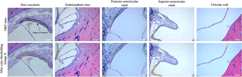
Methods: Mouse temporal bones were collected from juvenile (3-4 week) and aged (20-32 months) CBA/CaJ mice. Pediatric and adult human temporal bones from Caucasian or African-American individuals were examined from the Johns Hopkins Temporal Bone Collection. Information regarding Fitzpatrick skin type were unavailable, and self-identified race/ethnicity was used as a proxy. Images were taken using light microscopy at 20× magnification. ImageJ software (v1.53) was used to measure pigment within the SV and vestibular system.
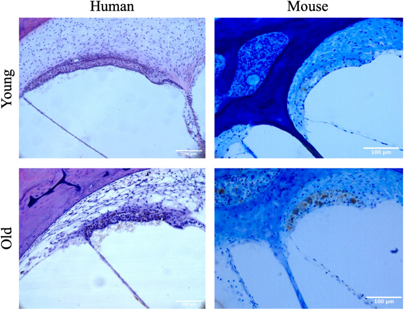
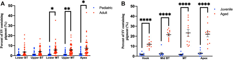
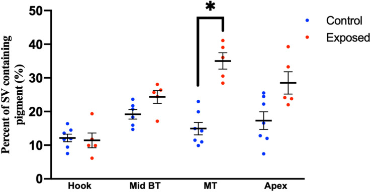
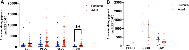
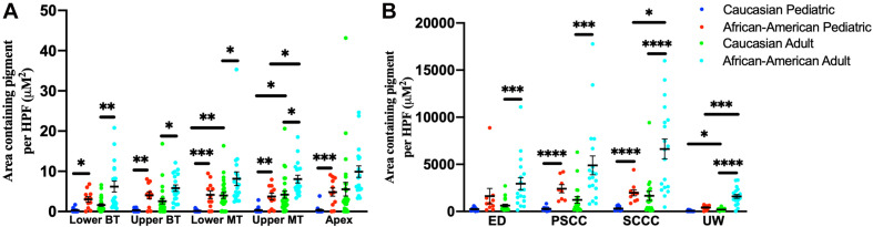
Results: In mouse temporal bones pigmentation within the SV increased with age, but pigmentation within the vestibular system did not increase with age. In human temporal bones pigmentation within the SV increased with age and pigmentation within the vestibular system increased within the wall of the utricle, but not other regions of the vestibular system. African-American individuals had higher amounts of pigment within the SV and vestibular system, among both pediatric and adult populations.
Conclusion: Stria vascularis pigmentation increases with age in mouse and human temporal bones. Pigmentation within the vestibular system did not increase with age in mouse specimens and only increased within the utricular wall with age in human specimens. Individuals who identified as African-American had higher pigment content within the SV and vestibular system, both as children and as adults. These results highlight how similar age-related pigmentary changes occur in the auditory and vestibular systems across species and underscore the importance of racial/ethnic diversity in human temporal bone studies.
Keywords: age-related hearing loss; auditory; cochlea; melanin; pigmentation; vestibular.
Copyright © 2021 Andresen, Coreas, Villavisanis and Lauer.
Conflict of interest statement
The authors declare that the research was conducted in the absence of any commercial or financial relationships that could be construed as a potential conflict of interest.
Figures
References
-
- Bonaccorsi P. (1965). [The color of the iris as a “test” in the quantitative estimation, in man, of the melanin concentration in the stria vascularis]. Ann. Laringol. Otol. Rinol. Faringol. 64 725–738. - PubMed
Grants and funding
LinkOut - more resources
Full Text Sources
Other Literature Sources