. 2021 May 12:12:671563.
doi: 10.3389/fphar.2021.671563. eCollection 2021.

Nicotinamide Riboside Enhances Endothelial Precursor Cell Function to Promote Refractory Wound Healing Through Mediating the Sirt1/AMPK Pathway

Affiliations

Nicotinamide Riboside Enhances Endothelial Precursor Cell Function to Promote Refractory Wound Healing Through Mediating the Sirt1/AMPK Pathway

Zhen-Hua Wang et al. Front Pharmacol. .

Abstract

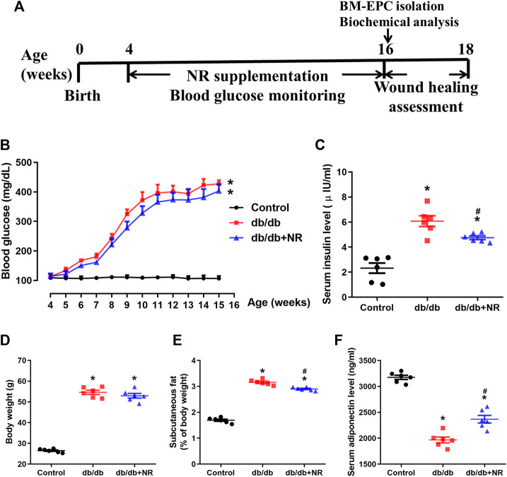
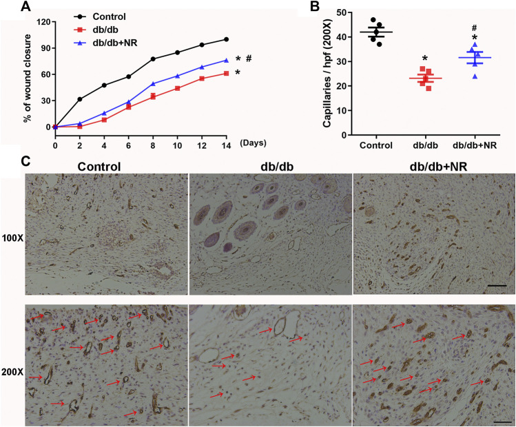
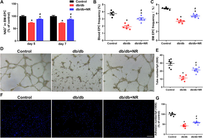
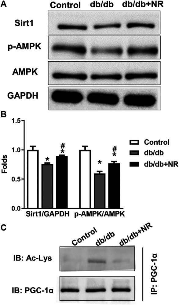
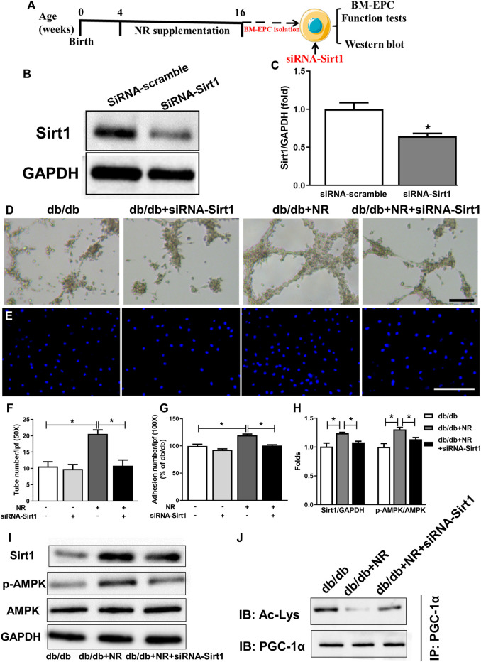
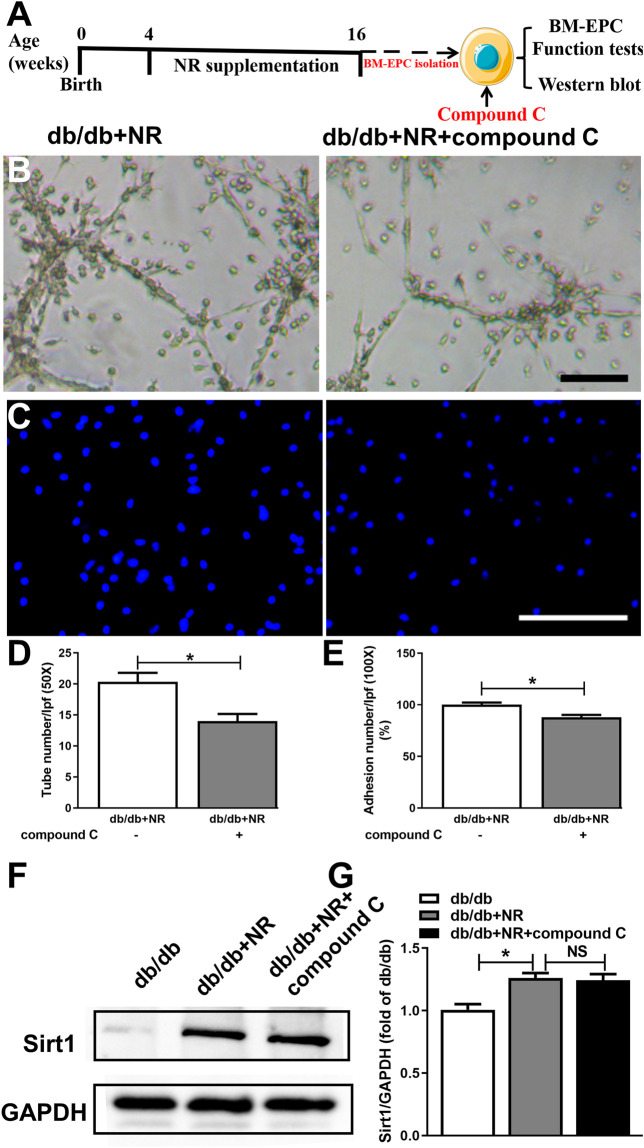
Lack of vascularization is directly associated with refractory wound healing in diabetes mellitus (DM). Enrichment of endothelial precursor cells (EPCs) is a promising but challenging approach for the treatment of diabetic wounds. Herein, we investigate the action of nicotinamide riboside (NR) on EPC function for improved healing of diabetic wounds. Db/db mice that were treated with NR-supplemented food (400 mg/kg/d) for 12 weeks exhibited higher wound healing rates and angiogenesis than untreated db/db mice. In agreement with this phenotype, NR supplementation significantly increased the number of blood EPCs and bone marrow (BM)-derived EPCs of db/db mice, as well as the tube formation and adhesion functions of BM-EPCs. Furthermore, NR-supplemented BM-EPCs showed higher expression of sirtuin 1 (Sirt1), phosphorylated adenosine monophosphate-activated protein kinase (p-AMPK), and lower expression of acetylated peroxisome proliferator-activated receptor γ coactivator (PGC-1α) than BM-EPCs isolated from untreated db/db mice. Knockdown of Sirt1 in BM-EPCs significantly abolished the tube formation and adhesion function of NR as well as the expression of p-AMPK and deacetylated PGC-1a. Inhibition of AMPK abolished the NR-regulated EPC function but had no effect on Sirt1 expression, demonstrating that NR enhances EPC function through the Sirt1-AMPK pathway. Overall, this study demonstrates that the oral uptake of NR enhances the EPC function to promote diabetic wound healing, indicating that NR supplementation might be a promising strategy to prevent the progression of diabetic complications.

Keywords: adenosine monophosphate–activated protein kinase; diabetes mellitus; endothelial precursor cells; nicotinamide riboside; sirtuin 1; wound healing.


Conflict of interest statement

The authors declare that the research was conducted in the absence of any commercial or financial relationships that could be construed as a potential conflict of interest.

Figures

FIGURE 1
FIGURE 1
Effects of NR supplement on some diabetes mellitus–related symptoms. (A) Experimental schedule. Db/db mice (4 weeks old) were fed with common food or NR-supplied food (400 mg/kg/d) for consecutive 12 weeks, and the blood glucose was monitored every week. After that, each group mice were further divided into 2 cohorts: excisional wound experiment and BM-EPC isolation experiment. Blood glucose (B), serum insulin (C), body weight (D), subcutaneous fat (E), and serum adiponectin (F) of the mice treated with common food or NR-supplied food for 12 weeks. Values are mean ± SEM, (n = 6). *P < 0.05 vs. control; # P < 0.05 vs. db/db. BM-EPC, bone marrow–derived endothelial precursor cell.
FIGURE 2
FIGURE 2
Effects of NR supplement on wound closure and wound angiogenesis in db/db mice. (A) Full-thickness skin wounds were made in common food and NR-supplied food fed db/db mice. Quantitative analysis of wound closure at indicated time intervals (B). Immunohistochemical analyses of CD31 in day-14 wounds of mice and quantitative study (C). Red arrows point out CD31-positive capillaries (100×, scale bar, 100 μm; 200×, scale bar, 50 μm). Values are mean ± SEM (n = 5). *P < 0.05 vs. control; # P < 0.05 vs. db/db.
FIGURE 3
FIGURE 3
Effects of NR supplement on EPC number and BM-EPC function in db/db mice. (A) NAD concentrations in cultured BM-EPCs (day 5 and day 7 in vitro) isolated from mice. Blood EPC (B) and BM EPC (C) frequency in normal control and db/db mice. Tube formation (D,E) and cell adhesion (F,G) of BM EPC isolated from normal control and db/db mice. D: 50×; scale bar, 100μm; F: 100×; scale bar, 100 μm. Values are mean ± SEM (A, B, C: n = 5; D, E, F, G: n = 6). *P < 0.05 vs. Control; # P < 0.05 vs. db/db. BM, bone marrow; BM-EPC, bone marrow–derived endothelial precursor cell.
FIGURE 4
FIGURE 4
Effect of NR supplement on the expression of Sirt1/AMPK in BM-EPCs. (A) Western blot of Sirt1/AMPK in BM-EPCs isolated from normal control and diabetic mice. (B) Quantitative analysis of Sirt1 and p-AMPK expression taken as in (A). (C) The level of acetylated PGC-1α was measured. Values are mean ± SEM, (n = 3). *P < 0.05 vs. Control; # P < 0.05 vs. db/db.
FIGURE 5
FIGURE 5
Inhibition of Sirt1 with siRNA-Sirt1 abolished the protective effects of NR repletion on BM-EPC function in db/db mice. (A) Experimental schedule. Db/db mice received the NR supplied food for 12 weeks, and then, BM-EPCs were isolated and transfected with siRNA targeting Sirt1. (B,C) Transfection of siRNA–Sirt1 obviously inhibited Sirt1 expression in BM-EPCs from db/db mice. *P < 0.05 vs. siRNA-Sirt1. Tube formation (D) and adhesion abilities of BM-EPCs (E) were measured. Quantitative analysis of tube number (F) and adhesion number (G). (H) Quantitative analysis of Sirt1, and p-AMPK expression was taken as in (I). (I) Western blot of Sirt1/AMPK in BM-EPCs treated with NR in the presence or absence of siRNA–Sirt1. (J) Representative immunoblot images of acetylated PGC-1α. D: 50×; scale bar, 100 μm; E: 100×; scale bar, 100 μm. Values are mean ± SEM, (B, C, H, I, J: n = 3; D–G: n = 5). *P < 0.05. BM-EPC, bone marrow–derived endothelial precursor cell; NS, no significance.
FIGURE 6
FIGURE 6
Inhibition of AMPK with compound C abolished the protective effects of NR supplement on BM-EPC function in db/db mice. (A) Illustration of experimental protocol. Db/db mice treated with NR for 12 weeks, and then, BM-EPCs were isolated and stimulated with compound C (10 μM). Tube formation (B) and adhesion (C) assay in BM-EPCs treated with NR in the presence or absence of compound C. Quantitated analysis of tube number (D) and adhesion number (E). (F–G) Compound C has no significant effect on Sirt1 expression in BM-EPCs of db/db mice treated with NR. B: 50×; scale bar, 100 μm; C: 100×; scale bar, 100 μm. Values are mean ± SEM (n = 3). *P < 0.05. BM-EPC, bone marrow–derived endothelial precursor cell; NS, no significance.

Similar articles

Cited by

References

    1. American Diabetes Association (2018). National Diabetes Statistics Report. Diabetes Care 2018, dci180007. 10.2337/dci18-0007 - DOI
    1. Azizi S., Kheirandish R., Salarpoor M. (2019). Topical Effect of Allogenous Serum Rich in Growth Factors (SRGF) on Diabetic Skin Wound in Rat. Transfus. Apher. Sci. 58 (4), 498–504. 10.1016/j.transci.2019.05.014 - DOI - PubMed
    1. Cantó C., Houtkooper R. H., Pirinen E., Youn D. Y., Oosterveer M. H., Cen Y., et al. (2012). The NAD+ Precursor Nicotinamide Riboside Enhances Oxidative Metabolism and Protects against High-Fat Diet-Induced Obesity. Cel Metab. 15 (6), 838–847. 10.1016/j.cmet.2012.04.022 - DOI - PMC - PubMed
    1. Carraway J. (2004). Using Aldara, Copper Peptide, and Niacinamide for Skin Care. Aesthet. Surg. J. 24 (1), 83–84. 10.1016/j.asj.2003.10.009 - DOI - PubMed
    1. Catrina S.-B., Zheng X. (2016). Disturbed Hypoxic Responses as a Pathogenic Mechanism of Diabetic Foot Ulcers. Diabetes Metab. Res. Rev. 32, 179–185. 10.1002/dmrr.2742 - DOI - PubMed

LinkOut - more resources