. 2021 May 14:12:636235.
doi: 10.3389/fneur.2021.636235. eCollection 2021.

Pre-operative MRI Radiomics for the Prediction of Progression and Recurrence in Meningiomas

Affiliations

Pre-operative MRI Radiomics for the Prediction of Progression and Recurrence in Meningiomas

Ching-Chung Ko et al. Front Neurol. .

Abstract

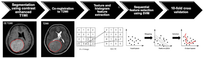
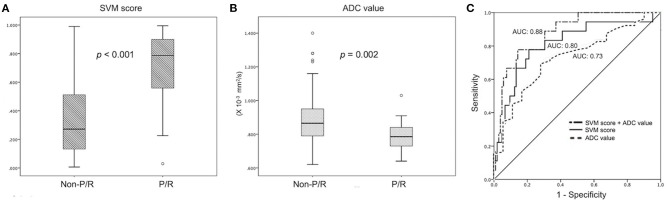
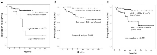
Objectives: A subset of meningiomas may show progression/recurrence (P/R) after surgical resection. This study applied pre-operative MR radiomics based on support vector machine (SVM) to predict P/R in meningiomas. Methods: From January 2007 to January 2018, 128 patients with pathologically confirmed WHO grade I meningiomas were included. Only patients who had undergone pre-operative MRIs and post-operative follow-up MRIs for more than 1 year were studied. Pre-operative T2WI and contrast-enhanced T1WI were analyzed. On each set of images, 32 first-order features and 75 textural features were extracted. The SVM classifier was utilized to evaluate the significance of extracted features, and the most significant four features were selected to calculate SVM score for each patient. Results: Gross total resection (Simpson grades I-III) was performed in 93 (93/128, 72.7%) patients, and 19 (19/128, 14.8%) patients had P/R after surgery. Subtotal tumor resection, bone invasion, low apparent diffusion coefficient (ADC) value, and high SVM score were more frequently encountered in the P/R group (p < 0.05). In multivariate Cox hazards analysis, bone invasion, ADC value, and SVM score were high-risk factors for P/R (p < 0.05) with hazard ratios of 7.31, 4.67, and 8.13, respectively. Using the SVM score, an AUC of 0.80 with optimal cutoff value of 0.224 was obtained for predicting P/R. Patients with higher SVM scores were associated with shorter progression-free survival (p = 0.003). Conclusions: Our preliminary results showed that pre-operative MR radiomic features may have the potential to offer valuable information in treatment planning for meningiomas.

Keywords: magnetic resonance imaging; meningioma; progression; radiomics; recurrence; support vector machine.


Conflict of interest statement

The authors declare that the research was conducted in the absence of any commercial or financial relationships that could be construed as a potential conflict of interest.

Figures

Figure 1
Figure 1
Flowchart indicating the process of analysis for the prediction of progression/recurrence (P/R) in meningiomas. The tumor is first segmented based on contrast-enhanced (CE) T1-weighted image (T1WI), and the region of interest (ROI) of the tumor is then mapped onto the T2-weighted image (T2WI). On each set of the two sequences, a total of 32 first-order features and 75 textural features are extracted, and a total of 214 parameters for each case are collected to develop the classification model. The most important four features are selected by means of the sequential feature selection and support vector machine (SVM) classifiers to calculate SVM score. The 10-fold cross-validation method is applied to test the model performance.
Figure 2
Figure 2
A 31-year-old woman with pathologically proven parafalcine meningioma (WHO grade I). (A) Axial CE T1WI showing an enhancing parafalcine tumor (red outline) at the frontal region. The tumor (red outline) is segmented on axial CE T1WI (A) and then mapped onto axial T2WI (B). The SVM score based on the four selected radiomic features is 0.831. (C) The measured ADC value (circular ROI) is 0.805 × 10⁻³ mm2/s (b = 1,000 s/mm2). (D) Gross total tumor resection is performed. (E,F) Progressive recurrence of tumor (arrowheads) was observed in 36 months (E) and 60 months (F) after surgery.
Figure 3
Figure 3
A 58-year-old man with pathologically proven parasagittal meningioma (WHO grade I). (A) Coronal CE T1WI shows an enhancing tumor mass (white arrow) in the midline parasagittal region with invasion into the superior sagittal sinus (SSS) (open black arrow) and adjacent skull bone (open curved arrow). (B,C) The tumor (red outline) is segmented on the axial CE T1WI (B) and then mapped onto the axial T2WI (C). Mild peritumoral edema (white open arrowheads) is noted on T2WI (C). The calculated SVM score based on the four selected radiomic features is 0.337. (D) Subtotal tumor resection is performed to preserve the SSS; residual tumor (curved arrow) is noted in the posterior SSS. (E,F) Progressive recurrence of tumor (white arrowheads) was observed in 37 months (E) and 56 months (F) after surgery.
Figure 4
Figure 4
Statistically significant differences (p < 0.05) (Mann–Whitney U test) are observed in the box plot of (A) SVM score and (B) ADC value to differentiate between patients with and without P/R. (C) Receiver operating characteristic (ROC) curves of SVM score and ADC value for the prediction of P/R in meningiomas, with optimal cutoff value of 0.224 and AUC of 0.825 × 10⁻³ mm2/s, respectively. The AUCs of SVM score, ADC value, and combination of SVM and ADC in the prediction of P/R are 0.80, 0.73, and 0.88, respectively.
Figure 5
Figure 5
Kaplan–Meier survival curves of (A) adjacent bone invasion, (B) SVM score, and (C) ADC value for the prediction of P/R in meningiomas. All three parameters showed significant difference (p < 0.05) (log-rank test) in overall trend of progression-free survival.

References

    1. Wiemels J, Wrensch M, Claus EB. Epidemiology and etiology of meningioma. J Neuro Oncol. (2010) 99:307–14. 10.1007/s11060-010-0386-3 - DOI - PMC - PubMed
    1. Louis DN, Perry A, Reifenberger G, von Deimling A, Figarella-Branger D, Cavenee WK, et al. . The 2016 world health organization classification of tumors of the central nervous system: a summary. Acta Neuropathol. (2016) 131:803–20. 10.1007/s00401-016-1545-1 - DOI - PubMed
    1. Maillo A, Orfao A, Espinosa AB, Sayagues JM, Merino M, Sousa P, et al. . Early recurrences in histologically benign/grade i meningiomas are associated with large tumors and coexistence of monosomy 14 and del(1p36) in the ancestral tumor cell clone. Neuro Oncol. (2007) 9:438–6. 10.1215/15228517-2007-026 - DOI - PMC - PubMed
    1. Perry A, Stafford SL, Scheithauer BW, Suman VJ, Lohse CM. Meningioma grading: an analysis of histologic parameters. Am J Surg Pathol. (1997) 21:1455–65. 10.1097/00000478-199712000-00008 - DOI - PubMed
    1. Ildan F, Erman T, Gocer AI, Tuna M, Bagdatoglu H, Cetinalp E, et al. . Predicting the probability of meningioma recurrence in the preoperative and early postoperative period: a multivariate analysis in the midterm follow-up. Skull Base. (2007) 17:157–71. 10.1055/s-2007-970554 - DOI - PMC - PubMed