Review
. 2021 May 14:12:661441.
doi: 10.3389/fneur.2021.661441. eCollection 2021.

Ulnar Neuropathy at the Elbow: From Ultrasound Scanning to Treatment

Affiliations
Review

Ulnar Neuropathy at the Elbow: From Ultrasound Scanning to Treatment

Kamal Mezian et al. Front Neurol. .

Abstract

Ulnar neuropathy at the elbow (UNE) is commonly encountered in clinical practice. It results from either static or dynamic compression of the ulnar nerve. While the retroepicondylar groove and its surrounding structures are quite superficial, the use of ultrasound (US) imaging is associated with the following advantages: (1) an excellent spatial resolution allows a detailed morphological assessment of the ulnar nerve and adjacent structures, (2) dynamic imaging represents the gold standard for assessing the ulnar nerve stability in the retroepicondylar groove during flexion/extension, and (3) US guidance bears the capability of increasing the accuracy and safety of injections. This review aims to illustrate the ulnar nerve's detailed anatomy at the elbow using cadaveric images to understand better both static and dynamic imaging of the ulnar nerve around the elbow. Pathologies covering ulnar nerve instability, idiopathic cubital tunnel syndrome, space-occupying lesions (e.g., ganglion, heterotopic ossification, aberrant veins, and anconeus epitrochlearis muscle) are presented. Additionally, the authors also exemplify the scientific evidence from the literature supporting the proposition that US guidance is beneficial in injection therapy of UNE. The non-surgical management description covers activity modifications, splinting, neuromobilization/gliding exercise, and physical agents. In the operative treatment description, an emphasis is put on two commonly used approaches-in situ decompression and anterior transpositions.

Keywords: US-guidance; cubital tunnel syndrome; elbow; entrapment neuropathy; musculoskeletal; peripheral nerve; ulnar nerve (MeSH); ultrasound.


Conflict of interest statement

The authors declare that the research was conducted in the absence of any commercial or financial relationships that could be construed as a potential conflict of interest.

Figures
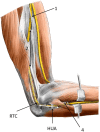
Figure 1
Figure 1
Possible sites of compression of the ulnar nerve at and around the elbow: (1) the medial intermuscular septum of the arm, (2) arcade of Struthers, (3) cubital tunnel, and (4) connective tissue between flexor carpi ulnaris and flexor digitorum superficialis muscles. RTC, retroepicondylar groove; HUA, humeroulnar aponeurotic arcade.
Figure 2
Figure 2
Course of the ulnar nerve in the arm. (A) Relationship of the ulnar nerve (U) and the medial septum (MIS). Ulnar nerve penetrates the medial septum—the penetration is demarcated by a ligamentous thickening (Struther's arcade). (B) Penetration of the septum and further course of the ulnar nerve is covered by muscular fibers of medial head of triceps brachii muscle, which begins on the medial intermuscular septum. (C) Transverse section of midarm depicting the relationship between ulnar nerve (U) and medial intermuscular septum (white arrow). BA, brachial artery; Bi, biceps brachii muscle; Br, brachialis muscle; BV, brachial vein; Hu, humerus; M, median nerve; ME, medial epicondyle; MIS, medial intermuscular septum; U, ulnar nerve; white arrow, medial intermuscular septum.
Figure 3
Figure 3
Retroepicondylar groove and the entrance to the cubital tunnel. (A) Entrance of the ulnar nerve to the cubital tunnel. (B) Demarcation of the cubital tunnel. BA, brachial artery; Br, brachialis muscle; BT, biceps brachii muscle tendon; BV, brachial vein; CFT, common flexor tendon; FCU, flexor carpi ulnaris muscle; ME, medial epicondyle; ole, olecranon; U, ulnar nerve; black arrow, medial collateral ligament, posterior bundle; white arrow, retroepicondylar retinaculum.
Figure 4
Figure 4
Cubital tunnel. (A) View on proximal part of the cubital tunnel with humeroulnar aponeurotic arcade (HUA) and ulnar nerve (U) going between humeral (1) and ulnar (2) head of flexor carpi ulnaris muscle (FCU). The course of ulnar nerve between FCU and flexor digitorum superficialis muscle (FDS). Humeral head is moved away, and the retroepicondylar retinaculum is transsected (white arrow). (B) Ulnar nerve exiting the cubital tunnel to forearm. The nerve is in close relationship to intermuscular connective tissue between FCU and FDS (black asterisk). (C) Cross section of the forearm depicting the relationship between the ulnar nerve and surrounding muscles and connective tissues (red and blue arrows). (1) flexor carpi ulnaris muscle, humeral head; (2) flexor carpi ulnaris muscle, ulnar head; HUA, humeroulnar aponeurotic arcade; Brachiorad, brachioradialis muscle; FCU, flexor carpi ulnaris muscle; FDP, flexor digitorum profundus muscle; FDS, flexor digitorum superficialis muscle; ole, olecranon; PrT, pronator teres; U, ulnar nerve; blue arrow, intermuscular connective tissue between FCU and FDS, deep flexor pronator aponeurosis; red arrow, intermuscular connective tissue between FCU and FDP.
Figure 5
Figure 5
Ultrasound (US) imaging of the ulnar nerve (U) at the level of the medial epicondyle (ME) in a patient with symptomatic ulnar nerve dislocation. Both longitudinal (A) and short-axis (B) views clearly demonstrate a close contact between the ulnar nerve and common flexor tendon origin. U, ulnar nerve; CFT, common flexor tendon; ME, medial epicondyle.
Figure 6
Figure 6
Normal ultrasound images of the ulnar nerve. (A) Short axis. (B) Long axis. U, ulnar nerve; ME, medial epicondyle; arrowheads, retroepicondylar retinaculum.
Figure 7
Figure 7
Proximal and distal tracking of the ulnar nerve starting from the retroepicondylar groove. (A) Midarm, arrowhead: medial intermuscular septum. (B) Proximal midarm. (C) Flexor carpi ulnaris muscle (FCU) level. (D) Proximal forearm. (E) Proximal mid-forearm, the ulnar nerve accompanied by the ulnar artery. (F) Ulnar nerve in Guyon canal (arrowheads). FDP, flexor digitorum profundus muscle; FDS, flexor digitorum superficialis muscle; U, ulnar nerve; BA, brachial artery; BV, brachial vein.
Figure 8
Figure 8
Comparative ultrasound (US) imaging of the ulnar nerve (U) at the level of the medial epicondyle (ME) in a patient with cubital tunnel syndrome. When compared with the normal side. (A) The asymptomatic side in a long axis of the ulnar nerve. (B) The symptomatic side ulnar nerve shows swelling (“bottle neck appearance”) proximal to the cubital tunnel inlet in long-axis. (D,E) In short axis, compared with the normal side (C), the ulnar nerve on the symptomatic side shows enlargement in its cross-sectional area of 18 mm2 outlined using the direct US tracing method (green dotted line). Hum, humerus; FCU, flexor carpi ulnaris muscle.
Figure 9
Figure 9
Ultrasound images of the ulnar nerve exemplary pathologies. (A) Short-axis image at the level of the humeral medial epicondyle (ME) shows the ulnar nerve (U) in an intimate contact with a ganglion (white arrowhead), likely derived from the triceps tendon. (B) A short-axis US image of the ulnar nerve situated just next to the heterotopic ossification (HO). (C) The ulnar nerve short-axis image shows an accessory anconeus epitrochlearis muscle (asterisk). (D) A longitudinal US image of the post-traumatic degenerative joint disease with effusion compressing the ulnar nerve. (E) A longitudinal image of the ulnar nerve depicts the nerve compression from scar tissue after olecranon surgery. (F) A short-axis view at the ulnar nerve (U) shows an aberrant vein (white arrow) next to it. ME, medial epicondyle of humerus; U, ulnar nerve.
Figure 10
Figure 10
(A) Prone position for the ultrasound-guided ulnar nerve in-plane injection with the elbow flexed and hanging over the examination bed. (B) The same procedure as described before in detail. (C) Supine position for the ultrasound-guided ulnar nerve in-plane injection; the patient is positioned on the examination bed with the elbow flexed and hand over the head. (D) The same procedure as described before in details.
Figure 11
Figure 11
(A) In situ decompression of the ulnar nerve in the cubital tunnel. (B) Anterior subcutaneous transposition—after wide decompression, the ulnar nerve is transposed anteriorly under the cutaneous flap.

References

    1. Staples JR, Calfee R. Cubital tunnel syndrome: current concepts. J Am Acad Orthop Surg. (2017) 25:e215–e24. 10.5435/JAAOS-D-15-00261 - DOI - PubMed
    1. Wu C-H, Chang K-V, Özçakar L, Hsiao M-Y, Hung C-Y, Shyu S-G, Wang T-G, et al. . Sonographic tracking of the upper limb peripheral nerves: a pictorial essay and video demonstration. Am J Phys Med Rehabil. (2015) 94:740–7. 10.1097/PHM.0000000000000344 - DOI - PubMed
    1. Kara M, Gürçay E, Ekiz T, Sekizkardeş M, Yorulmaz E, Ata AM, et al. . EURO-MUSCULUS/USPRM global report on musculoskeletal ultrasound publications. Am J Phys Med Rehabil. (2020) 99:847–52. 10.1097/PHM.0000000000001390 - DOI - PubMed
    1. Ricci V, Özçakar L. Ultrasonographic examination: the “X-Factor” in physiatry. Am J Phys Med Rehabil. (2020) 99:e131–e2. 10.1097/PHM.0000000000001372 - DOI - PubMed
    1. Mezian K, Steyeorova P, Vacek J, Navratil L. Introduction to neuromuscular ultrasound. Cesk Slov Neurol N. (2016) 79:656–61. 10.14735/amcsnn2016656 - DOI

LinkOut - more resources