Review
. 2021 May 14:12:675419.
doi: 10.3389/fmicb.2021.675419. eCollection 2021.

Therapeutic Potential of Exploiting Autophagy Cascade Against Coronavirus Infection

Affiliations
Review

Therapeutic Potential of Exploiting Autophagy Cascade Against Coronavirus Infection

Subhajit Maity et al. Front Microbiol. .

Abstract

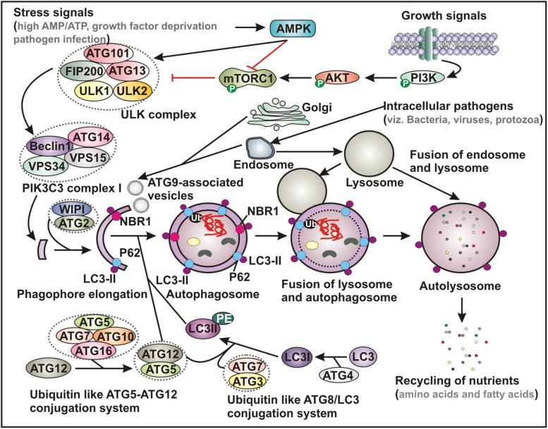
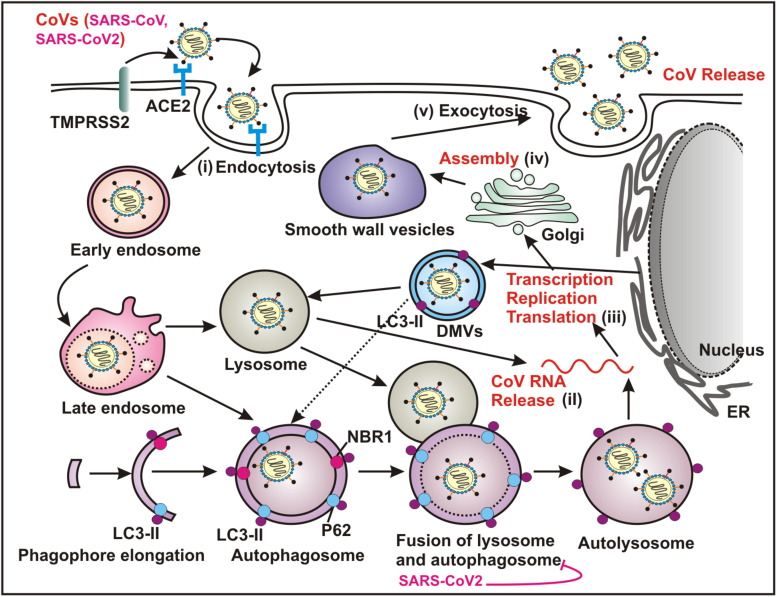
Since its emergence in December 2019 in Wuhan, China, severe acute respiratory syndrome coronavirus 2 (SARS-CoV-2) created a worldwide pandemic of coronavirus disease (COVID-19) with nearly 136 million cases and approximately 3 million deaths. Recent studies indicate that like other coronaviruses, SARS-CoV-2 also hijacks or usurps various host cell machineries including autophagy for its replication and disease pathogenesis. Double membrane vesicles generated during initiation of autophagy cascade act as a scaffold for the assembly of viral replication complexes and facilitate RNA synthesis. The use of autophagy inhibitors - chloroquine and hydroxychloroquine initially appeared to be as a potential treatment strategy of COVID-19 patients but later remained at the center of debate due to high cytotoxic effects. In the absence of a specific drug or vaccine, there is an urgent need for a safe, potent as well as affordable drug to control the disease spread. Given the intricate connection between autophagy machinery and viral pathogenesis, the question arises whether targeting autophagy pathway might show a path to fight against SARS-CoV-2 infection. In this review we will discuss about our current knowledge linking autophagy to coronaviruses and how that is being utilized to repurpose autophagy modulators as potential COVID-19 treatment.

Keywords: COVID-19; SARS-CoV-2; autophagy; coronaviruses (CoVs); virophagy.


Conflict of interest statement

The authors declare that the research was conducted in the absence of any commercial or financial relationships that could be construed as a potential conflict of interest.

Figures

FIGURE 1
FIGURE 1
Schematic representation of basic autophagy pathway. Upon deprivation of growth factors, increase in AMP level and pathogen infection lead to AMPK activation and subsequent inhibition of mTORC1 function. In contrast, in the presence of growth signals PI3K-AKT signaling pathway activates mTOR. Inhibition of mTORC1 results in activation of ULK complex, which phosphorylates Beclin-1, leading to VPS34 activation and initiation of phagophore formation. ULK functions in a complex with ULK1, ULK2, FIP200, ATG13 and ATG101, while VPS34 function within the PIK3C3 complex containing its regulatory subunit, VPS15, ATG14 and Beclin-1, which further recruits to WIPI and ATG2 for phagophore elongation. Several ATG proteins engage two evolutionarily conserved ubiquitin-like conjugation systems - ATG12-ATG5 and phosphatidylethanolamine (PE)-conjugated LC3 (LC3-II) targeted to the pre-autophagosomal membrane. In the ATG12-ATG5 conjugation system, the complex further interacts with ATG16, where ATG7 functions as an E1-like enzyme and ATG10 factions as an E2-like enzyme. In the other system, LC3 is first cleaved by a cysteine protease ATG4 to generate LC3-I, which is further conjugated with PE to form membrane bound LC3-II facilitated by ATG7 and ATG3. The cytoplasmic damaged cargo is then ubiquitinated, captured by adaptor molecules - p62 or NBR1 and subsequently delivered to the phagophore membrane. Matured autophagosome then fuses with endolysosomal vesicles forming an autolysosome, where the cargo is degraded and provide nutrients. AMPK, AMP activated protein kinase; mTORC1, mammalian target of rapamycin complex 1; PI3K, phosphatidylinositol 3-kinase; PIK3C3, Phosphatidylinositol 3-Kinase Catalytic Subunit Type 3; LC3, microtubule-associated protein 1 light chain 3; Ub, ubiquitin; NBR1, neighbor of BRCA1 gene 1.
FIGURE 2
FIGURE 2
Coronaviruses (CoVs) utilize autophagy pathway for replication. Both SARS-CoV and SARS-COV-2 recognize ACE2 as the cellular surface receptor that mediates the viral entry into the host via endocytosis. Several CoVs utilize ATG5/ATG7 independent autophagy that establishes an endocytic pathway-Golgi route potentially interacting with the multiple stages of virus replication cycle - (i) entry mechanism by endocytosis and fusion of viral and host membranes; (ii) uncoating and releasing the viral genomic RNA; (iii) transcription, replication and translation; (iv) trafficking and assembly and (v) egress by exocytosis. The replication transcription centers are intimately associated with DMVs, which are generated from the ER. DMVs mimic autophagosomes and subsequently these structures fuse with the late endosome, and the lysosome, which results in degradation of the sequestered cytoplasmic cargo. A number of CoVs including SARS-CoV-2 specifically block the fusion between autophagosome and lysosome. ACE2, angiotensin converting enzyme 2; ER, endoplasmic reticulum; DMVs, double membrane vesicles.

Similar articles

Cited by

References

    1. Ahmad L., Mostowy S., Sancho-Shimizu V. (2018). Autophagy-virus interplay: from cell biology to human disease. Front. Cell. Dev. Biol. 6:155. 10.3389/fcell.2018.00155 - DOI - PMC - PubMed
    1. Albert S., Serova M., Dreyer C., Sablin M. P., Faivre S., Raymond E. (2010). New inhibitors of the mammalian target of rapamycin signaling pathway for cancer. Expert. Opin. Invest. Drugs 19 919–930. - PubMed
    1. An H., Harper J. W. (2018). Systematic analysis of ribophagy in human cells reveals bystander flux during selective autophagy. Nat. Cell. Biol. 20 135–143. 10.1038/s41556-017-0007-x - DOI - PMC - PubMed
    1. Barnes C. O., West A. P., Jr., Huey-Tubman K. E., Hoffmann M. A. G., Sharaf N. G., Hoffman P. R., et al. (2020). Structures of human antibodies bound to SARS-CoV-2 spike reveal common epitopes and recurrent features of antibodies. Cell 182 828–842. 10.1016/j.cell.2020.06.025 - DOI - PMC - PubMed
    1. Beigel J. H., Tomashek K. M., Dodd L. E., Mehta A. K., Zingman B. S., Kalil A. C., et al. (2020). Remdesivir for the treatment of covid-19 - final report. N. Engl. J. Med. 383 1813–1826. - PMC - PubMed

LinkOut - more resources