. 2021 May 12:12:606963.
doi: 10.3389/fimmu.2021.606963. eCollection 2021.

Autoantibodies Against Ubiquitous and Confined Antigens in Patients With Ocular, Neuro-Ophthalmic and Congenital Cerebral Toxoplasmosis

Affiliations

Autoantibodies Against Ubiquitous and Confined Antigens in Patients With Ocular, Neuro-Ophthalmic and Congenital Cerebral Toxoplasmosis

Monica Goldberg-Murow et al. Front Immunol. .

Abstract

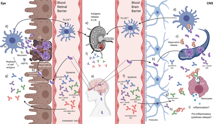
Toxoplasma gondii infection can trigger autoreactivity by different mechanisms. In the case of ocular toxoplasmosis, disruption of the blood-retinal barrier may cause exposure of confined retinal antigens such as recoverin. Besides, cross-reactivity can be induced by molecular mimicry of parasite antigens like HSP70, which shares 76% identity with the human ortholog. Autoreactivity can be a determining factor of clinical manifestations in the eye and in the central nervous system. We performed a prospective observational study to determine the presence of autoantibodies against recoverin and HSP70 by indirect ELISA in the serum of 65 patients with ocular, neuro-ophthalmic and congenital cerebral toxoplasmosis. We found systemic autoantibodies against recoverin and HSP70 in 33.8% and 15.6% of individuals, respectively. The presence of autoantibodies in cases of OT may be related to the severity of clinical manifestations, while in cases with CNS involvement they may have a protective role. Unexpectedly, anti-recoverin antibodies were found in patients with cerebral involvement, without ocular toxoplasmosis; therefore, we analyzed and proved cross-reactivity between recoverin and a brain antigen, hippocalcin, so the immunological phenomenon occurring in one immune-privileged organ (e.g. the central nervous system) could affect the environment of another (egg. the eye).

Keywords: HSP70; Toxoplasma gondii; autoantibodies; cerebral toxoplasmosis; cross-reactivity; hippocalcin; ocular toxoplasmosis; recoverin.


Conflict of interest statement

The authors declare that the research was conducted in the absence of any commercial or financial relationships that could be construed as a potential conflict of interest.

Figures

Figure 1
Figure 1
Sequence alignment of the Homo sapiens (upper rows) and T. gondii HSP70s (lower rows). In the middle lines are the matching amino acids. The identity between the sequences was 76% (yellow circle), in lilac and green colors are the B antigenic determinants predicted for the respective sequences. The arrows indicate the antigenic determinants that both proteins share.
Figure 2
Figure 2
Presence of anti-HSP70 (upper graph) and anti-recoverin (lower graph) autoantibodies in the different clinical groups; the dispersion of the reactivity indices to HSP70 and recoverin is shown, as well as the differences among them (analyzed with ∞ Mann-Whitney U and κ Kruskal-Wallis.) and the proportion of positives RI > 1.0 (analyzed with Φ Fisher’s exact test). The only statistically significant result obtained was the difference between positives in the isolated OT (acquired) and the subclinical group).
Figure 3
Figure 3
Correlation between the RI to HSP70 and RI to recoverin of the positive samples to one or both antigens. ς Spearman’s correlation.
Figure 4
Figure 4
Response to HSP70 (upper graph) and recoverin (lower graph) in the subgroup of neuro-ophthalmic toxoplasmosis. P values using ∞ Mann-Whitney U test, or κ Kruskal-Wallis.
Figure 5
Figure 5
Response to recoverin in patients with congenital cerebral toxoplasmosis without OT. ∞ Mann-Whitney U test.
Figure 6
Figure 6
Alignment of hippocalcin (upper row) and recoverin (lower row) sequences. In the middle lines are the matching amino acids. Circulated in yellow is the identity between the sequences, and in the orange and red boxes are the B epitopes predicted in each sequence. The arrows indicate the antigenic determinants that the proteins share.
Figure 7
Figure 7
Presence of anti-hippocalcin autoantibodies in the different clinical groups. The graph shows the hippocalcin reactivity of the selected samples and the proportion of positives, i.e. RI ≥ 1.0. Statistics performed with Kruskal-Wallis.
Figure 8
Figure 8
Immunochemical analysis of the anti-recoverin and anti-hippocalcin responses. Correlation between absorbance and avidity for anti-recoverin and anti-hippocalcin. Correlation between absorbance and avidity values to recoverin and hippocalcin. Spearman’s correlation test.
Figure 9
Figure 9
Recoverin-hippocalcin inhibition assay. For each patient the avidity to recoverin and hippocalcin is shown on the left side, the percent inhibition is plotted on the right side according to the added concentration of the competing antigen under four different conditions: – R + R plate coated with recoverin + serum treated with recoverin; - - - R + H plate coated with recoverin + serum treated with hippocalcin; – H + H plate coated with hippocalcin + serum treated with hippocalcin, - - - H + R plate coated with hippocalcin + serum treated with recoverin.
Figure 10
Figure 10
T. gondii induces the production of autoantibodies that might be related to disease severity in CNS or the eye. a) T. gondii tachyzoites can migrate throughout the blood to reach the CNS and the retina, b) infecting local cells. c) Tachyzoites replication and exit promote lysis, and endogen antigens are released from damaged tissues. d) Tissue-resident DCs could recognize and engulf T. gondii tachyzoites and endogen antigens. e) DCs activation can promote its mobilization to the closest lymphatic node (LN) to present antigens, f) probably activating B cells to produce anti-T. gondii and anti-endogen antigens antibodies that can be detected systemically. g) These antibodies could reach the retina and the CNS. h) Autoantibodies against recoverin show cross-reactivity against other self-antigens such as hippocalcin, i) which in CNS might be related to increasing a pro-inflammatory environment beneficial against the parasite. Created with BioRender.com.

References

    1. Rosenblum MD, Remedios KA, Abbas AK. Mechanisms of Human Autoimmunity. J Clin Invest (2015) 125:2228–33. 10.1172/JCI78088 - DOI - PMC - PubMed
    1. Kivity S, Agmon-Levin N, Blank M, Shoenfeld Y. Infections and Autoimmunity – Friends or Foes? Trends Immunol (2009) 30:409–14. 10.1016/j.it.2009.05.005 - DOI - PubMed
    1. Montoya J, Liesenfeld O. Toxoplasmosis. Lancet (2004) 363:1965–76. 10.1016/S0140-6736(04)16412-X - DOI - PubMed
    1. Suzuki Y, Sa Q, Ochiai E, Mullins J, Yolken R, Halone S. Cerebral Toxoplasmosis: Pathogenesis and Host Resistance. In: Weiss LM, Kim K, editors. Toxoplasma gondii: The Model Apicomplexan: Perspectives and Methods. London: Academic Press; (2013). p. 567–91.
    1. Butler NJ, Furtado JM, Winthrop KL, Smith JR. Ocular Toxoplasmosis II: Clinical Features, Pathology and Management. Clin Exp Ophthalmol (2013) 41:95–108. 10.1111/j.1442-9071.2012.02838.x - DOI - PMC - PubMed

Publication types

MeSH terms

LinkOut - more resources