. 2021 May 13:12:613438.
doi: 10.3389/fimmu.2021.613438. eCollection 2021.

Latent TGF-β Activation Is a Hallmark of the Tenascin Family

Affiliations

Latent TGF-β Activation Is a Hallmark of the Tenascin Family

Alexandre Aubert et al. Front Immunol. .

Abstract

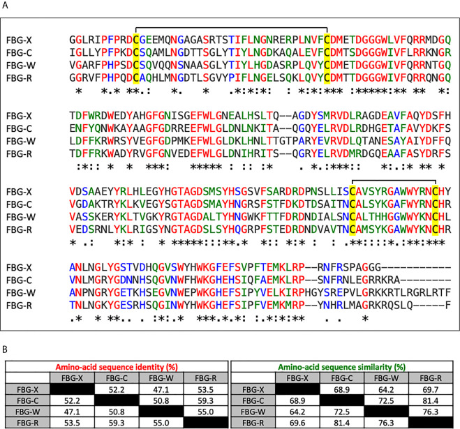
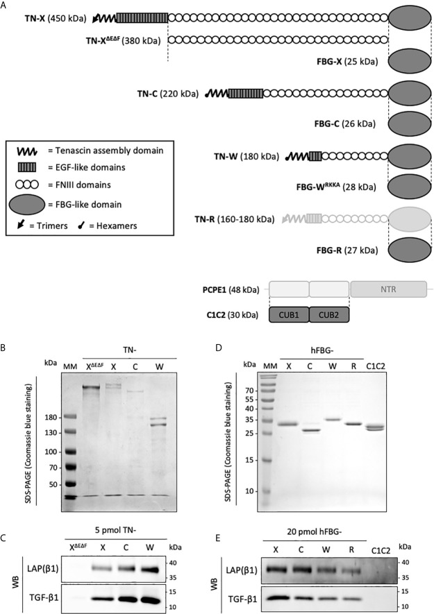
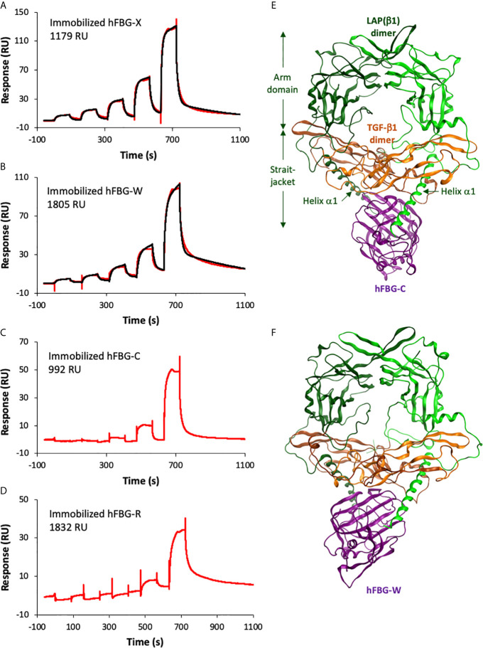
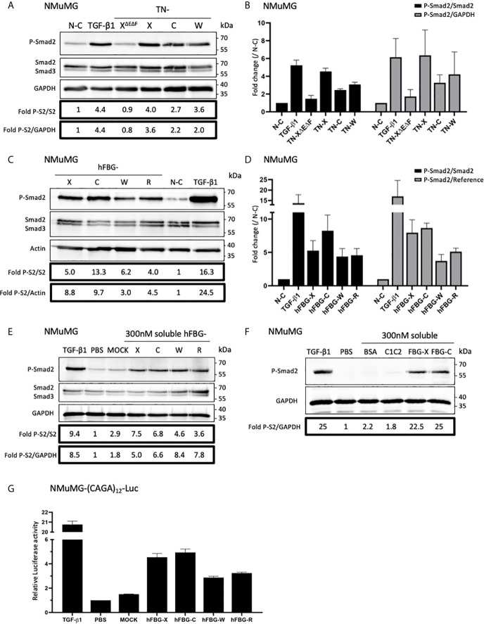
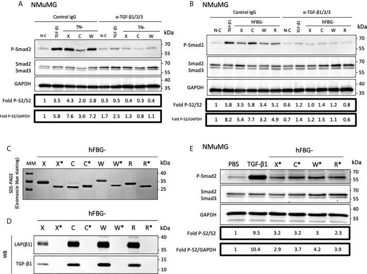
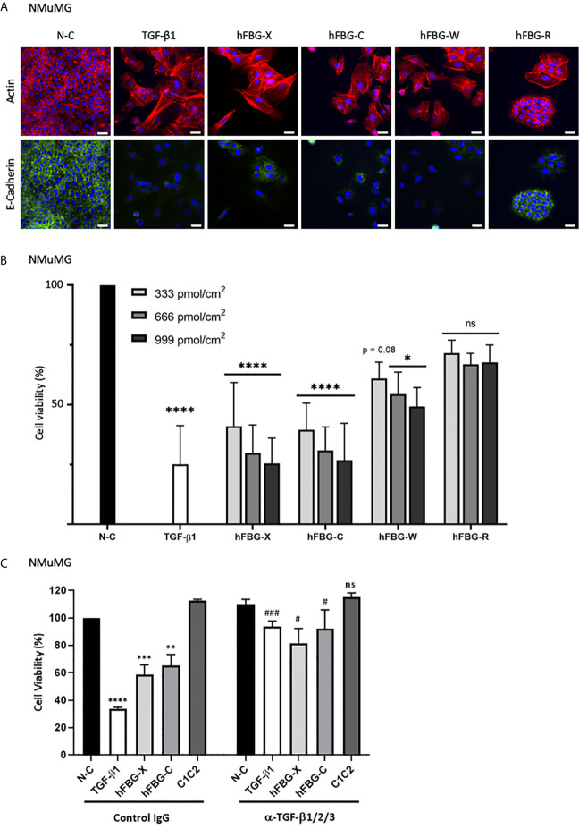
Transforming growth factor-β (TGF-β) isoforms are secreted as inactive complexes formed through non-covalent interactions between bioactive TGF-β entities and their N-terminal pro-domains called latency-associated peptides (LAP). Extracellular activation of latent TGF-β within this complex is a crucial step in the regulation of TGF-β activity for tissue homeostasis and immune cell function. We previously showed that the matrix glycoprotein Tenascin-X (TN-X) interacted with the small latent TGF-β complex and triggered the activation of the latent cytokine into a bioactive TGF-β. This activation most likely occurs through a conformational change within the latent TGF-β complex and requires the C-terminal fibrinogen-like (FBG) domain of the glycoprotein. As the FBG-like domain is highly conserved among the Tenascin family members, we hypothesized that Tenascin-C (TN-C), Tenascin-R (TN-R) and Tenascin-W (TN-W) might share with TN-X the ability to regulate TGF-β bioavailability through their C-terminal domain. Here, we demonstrate that purified recombinant full-length Tenascins associate with the small latent TGF-β complex through their FBG-like domains. This association promotes activation of the latent cytokine and subsequent TGF-β cell responses in mammary epithelial cells, such as cytostasis and epithelial-to-mesenchymal transition (EMT). Considering the pleiotropic role of TGF-β in numerous physiological and pathological contexts, our data indicate a novel common function for the Tenascin family in the regulation of tissue homeostasis under healthy and pathological conditions.

Keywords: immune cell modulation; latent TGF-β activation; tenascins; tissue homeostasis; transforming growth factor (TGF)-β; tumor microenvironment.


Conflict of interest statement

The authors declare that the research was conducted in the absence of any commercial or financial relationships that could be construed as a potential conflict of interest.

Figures

Figure 1
Figure 1
Sequence alignment and homology degree between the Tenascin family FBG-like domains. (A) Multiple sequence alignment analysis of the FBG-like domains of human TN-X, -C, -W and -R (FBG-X, -C, -W, -R) performed with CLUSTALW software. Red (*) labelled residues are identical between the four FBG-like domains, green labelled (:) residues are highly conserved and blue labelled (.) residues are poorly conserved. The four conserved cysteine residues are highlighted in yellow. (B) Percentages of identity and similarity amongst the amino-acid sequences of the FBG-like domains of the human Tenascins performed with paired comparisons using CLUSTALW software.
Figure 2
Figure 2
Recombinant full-length Tenascins and their respective FBG-like domains co-purify with latent TGF-β1. (A) Schematic representation of the recombinant proteins used in this study (dark grey). Structural modules of Tenascins are depicted in the inset. (B, D) SDS-PAGE analysis of the purified recombinant proteins and stained with Coomassie blue. MM, molecular mass markers. (B) Purified full-length TN-X, TN-C, TN-W and the TN-XΔEΔF fragment (1 μg each) were loaded on 6% acrylamide gels under reducing conditions. (D) Purified recombinant FBG-like domains of the four human Tenascins and the recombinant CUB1CUB2 (C1C2) domain (2 μg) were resolved on 15% acrylamide gels under reducing conditions. (C, E) Western blot analysis indicating the level of human mature TGF-β1 and LAP(β1) pro-domain associated with equimolar quantity of purified recombinant full-length TN or TN-XΔEΔF fragment (5 pmol each) (C) and recombinant Tenascin FBG-like domains or CUB1CUB2 (C1C2) protein (20 pmol) (E).
Figure 3
Figure 3
The recombinant FBG-like domains of Tenascins interact with TGF-β1. (A–D) Interactions of TGF-β1 with FBG-like domains analyzed by SPR. Increasing concentrations of TGF-β1 were injected in single-cycle mode over immobilized FBG-X (A), FBG-W (B), FBG-C (C) and FBG-R (D). The experimental curves are shown in red and the best fits with the heterogeneous ligand model in black. The concentrations of TGF-β1 were as follows: (A) 15.6 – 31.2 – 62.5 – 125 – 250 nM; (B) 7.8 – 15.6 – 31.25 – 62.5 – 125 nM; (C) 20.2 – 40.5 – 81 – 162 – 324 nM; (D) 9.4 – 18.8 – 37.5 – 75 – 150 nM. (E, F) Homology models showing the predicted docking between the FBG-like domain of TN-C (E) or TN-W (F) and the pro-TGF-β1. The dimeric LAP(β1) pro-domain is shown in green, the TGF-β1 dimer in orange and the FBG-like domains in purple. ‘Straitjacket’ and arm domains of the LAP(β1) dimer are indicated.
Figure 4
Figure 4
Full-length Tenascins and their respective FBG-like domains stimulate TGF-β/Smad intracellular signaling pathway in epithelial cells. (A) Western Blot analysis showing phosphorylated Smad2 (P-Smad2), total Smad2/3 and GAPDH levels in NMuMG cells cultured for 3h onto control non-coated (N-C) dishes or dishes coated with equimolar quantity (22,2 pmol/cm2) of full-length Tenascins (TN) or central TN-X fragment (TN-XΔEΔF), or stimulated with soluble active TGF-β1 (2 ng/mL). (B) Fold changes of P-Smad2 to total Smad2 levels or to GAPDH levels. Graph shows means ± SD of n = 3 independent experiments. (C) Western Blot analysis showing P-Smad2, total Smad2/3 and Actin levels in NMuMG cultured for 3h onto equimolar quantity of FBG-like domains (333 pmol/cm2) or stimulated with soluble active TGF-β1 (2 ng/mL). (D) Fold changes of P-Smad2 to total Smad2 levels or to reference protein levels. Graph shows means ± SD of n = 3 independent experiments. (E) Western-blot analysis showing P-Smad2, Smad2/3 and GAPDH levels in NMuMG cells stimulated for 1h in the presence of active TGF-β1 (2 ng/mL), stimulated or not (PBS) with soluble FBG-like domains (300 nM) or MOCK-CM. “MOCK-CM” referred to the conditioned media of MOCK-transfected HEK293 EBNA cells, submitted to Nickel-affinity chromatography and eluted from the column as for the FBG-like domains. (F) Immunoblotting analysis showing P-Smad2 and GAPDH levels in NMuMG cells stimulated for 1h in the presence of active TGF-β1 (2 ng/mL), stimulated or not (PBS) with soluble CUB1CUB2 protein fragment (C1C2), bovine serum Albumin (BSA) or FBG-like domains (300 nM each). (G) Relative Luciferase activity of NMuMG cells transiently transfected with the Smad-responsive (CAGA)12-Luc reporter construct and treated for 16h with soluble FBG-like domains (600 nM), MOCK-CM or TGF-β1 (2 ng/mL). Graph shows one representative result from 3 independent experiments.
Figure 5
Figure 5
The FBG-like domain of the Tenascin family members activates the latent TGF-β in epithelial cells. (A) Western-blot analysis showing P-Smad2, total Smad2/3 and GAPDH levels in NMuMG cultured for 3h onto Non-Coated (N-C) dishes, dishes coated with 22,2 pmol/cm2 full-length Tenascins (TN) or stimulated with 2 ng/mL TGF-β1, in presence of anti-pan-TGF-β antibody or isotype-matched control IgG (10 μg/mL). (B) Western Blot analysis showing P-Smad2, total Smad2/3 and GAPDH levels in NMuMG cells cultured for 3h in presence of 333 pmol/cm2 of FBG-like domains as described in (A). (C) SDS-PAGE analysis of purified recombinant FBG-like domains produced in mammalian cells (FBG) and E. coli (FBG*) resolved on 15% acrylamide gels under reducing conditions (2 µg each). MM, molecular mass markers. (D) Western-Blot analysis of the levels of mature TGF-β1 and LAP(β1) pro-domain associated with 20 pmol of FBG-like domains produced in mammalian cells (FBG) and E coli (FBG*). (E) Western-blot analysis showing P-Smad2, Smad2/3 and GAPDH levels in NMuMG cells stimulated for 1h with active TGF-β1 (2 ng/mL), soluble purified FBG-like domains produced in E Coli (720 nM) or vehicle (PBS).
Figure 6
Figure 6
The FBG-like domains of Tenascins trigger EMT and cytostasis in epithelial cells. (A) F-actin direct fluorescence (red) and E-Cadherin indirect immunofluorescence (green) performed on NMuMG cells cultured for 72h onto N-C dishes, dishes coated with 666 pmol/cm2 FBG-like domains or stimulated with soluble TGF-β1 (5 ng/mL). Cell nuclei were counterstained with DAPI (blue). Bars, 15 µm. (B) Percentage of cell viability in NMuMG cells cultured for 48h onto N-C dishes, dishes coated with 333, 666 or 999 pmol/cm2 FBG-like domains, or stimulated with 5 ng/mL TGF-β1. Error bars are means ± SD from 3 independent experiments. *p < 0.05 compared to N-C condition. ****p < 0.0001 compared to N-C condition. ns, not significant. (C) Percentage of cell viability in NMuMG cells cultured for 48h onto N-C dishes, dishes coated with 666 pmol/cm2 FBG-like domains (FBG-X and FBG-C) or CUB1CUB2 protein fragment (C1C2), or stimulated with 5 ng/mL TGF-β1, in the presence of anti-pan-TGF-β antibody or isotype-matched control IgG (5 μg/mL). Error bars are means ± SD from 3 independent experiments. **, *** and **** respectively correspond to p < 0.01, p < 0.001 and p < 0.0001 compared to N-C condition. # and ### respectively correspond to p < 0.05 and p < 0.001 versus their control IgG-treated counterpart. ns, not significant.

References

    1. Chiquet-Ehrismann R, Tucker RP. Tenascins and the Importance of Adhesion Modulation. Cold Spring Harb Perspect Biol (2011) 3(5):a004960. 10.1101/cshperspect.a004960 - DOI - PMC - PubMed
    1. Nörenberg U, Wille H, Wolff JM, Frank R, Rathjen FG. The Chicken Neural Extracellular Matrix Molecule Restrictin: Similarity With EGF-, Fibronectin Type III-, and Fibrinogen-Like Motifs. Neuron (1992) 8:849–63. 10.1016/0896-6273(92)90199-N - DOI - PubMed
    1. Lethias C, Carisey A, Comte J, Cluzel C. Exposito J-Y. A Model of Tenascin-X Integration Within the Collagenous Network. FEBS Lett (2006) 580:6281–5. 10.1016/j.febslet.2006.10.037 - DOI - PubMed
    1. Erickson HP, Taylor HC. Hexabrachion Proteins in Embryonic Chicken Tissues and Human Tumors. J Cell Biol (1987) 105:1387–94. 10.1083/jcb.105.3.1387 - DOI - PMC - PubMed
    1. Scherberich A, Tucker RP, Samandari E, Brown-Luedi M, Martin D, Chiquet-Ehrismann R. Murine tenascin-W: A Novel Mammalian Tenascin Expressed in Kidney and At Sites of Bone and Smooth Muscle Development. J Cell Sci (2004) 117:571–81. 10.1242/jcs.00867 - DOI - PubMed

Publication types

MeSH terms