. 2021 May 12:12:622935.
doi: 10.3389/fimmu.2021.622935. eCollection 2021.

Intra-Abdominal Lipopolysaccharide Clearance and Inactivation in Peritonitis: Key Roles for Lipoproteins and the Phospholipid Transfer Protein

Affiliations

Intra-Abdominal Lipopolysaccharide Clearance and Inactivation in Peritonitis: Key Roles for Lipoproteins and the Phospholipid Transfer Protein

Maxime Nguyen et al. Front Immunol. .

Abstract

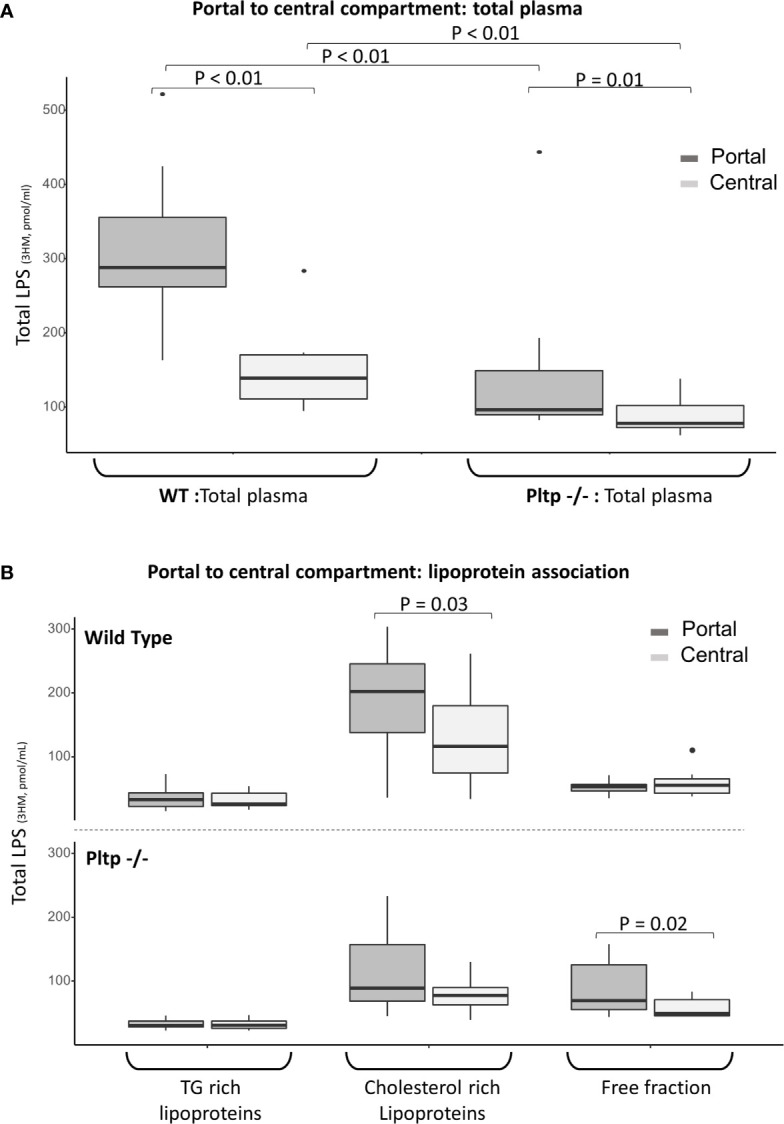
Introduction: During peritonitis, lipopolysaccharides (LPS) cross the peritoneum and pass through the liver before reaching the central compartment. The aim of the present study was to investigate the role of lipoproteins and phospholipid transfer protein (PLTP) in the early stages of LPS detoxification.

Material and methods: Peritonitis was induced by intra-peritoneal injection of LPS in mice. We analyzed peritoneal fluid, portal and central blood. Lipoprotein fractions were obtained by ultracentrifugation and fast protein liquid chromatography. LPS concentration and activity were measured by liquid chromatography coupled with mass spectrometry and limulus amoebocyte lysate. Wild-type mice were compared to mice knocked out for PLTP.

Results: In mice expressing PLTP, LPS was able to bind to HDL in the peritoneal compartment, and this was maintained in plasma from portal and central blood. A hepatic first-pass effect of HDL-bound LPS was observed in wild-type mice. LPS binding to HDL resulted in an early arrival of inactive LPS in the central blood of wild-type mice.

Conclusion: PLTP promotes LPS peritoneal clearance and neutralization in a model of peritonitis. This mechanism involves the early binding of LPS to lipoproteins inside the peritoneal cavity, which promotes LPS translocation through the peritoneum and its uptake by the liver.

Keywords: endotoxemia; inflammation; lipopolysaccharides 4; lipoproteins; peritonitis; phospholipid transfer protein; sepsis.


Conflict of interest statement

The authors declare that the research was conducted in the absence of any commercial or financial relationships that could be construed as a potential conflict of interest.

Figures

Figure 1
Figure 1
Characterization of lipoproteins in central, portal, and peritoneal compartments (from pooled samples) in wild-type (black lines) and Pltp -/- (grey lines) mice in absence of LPS injections by (A) Electrophoresis and (B) Fast protein liquid chromatography. Both electrophoresis and FPLC showed clear differences in lipoprotein composition between portal and central blood. For FPLC, fractions 5 to 37 are shown. FPLC, Fast protein liquid chromatography; Pltp -/-, Knock out for the phospholipid transfer protein; VLDL, very low density lipoprotein; LDL, low density lipoprotein; HDL, High density lipoprotein.
Figure 2
Figure 2
Kinetics of central LPS concentration and activity in wild-type (black lines) and Pltp -/- (grey lines) mice. (A) After intra-peritoneal injection (n = 8 vs. n = 7) and (B) after caudal (intravenous) injection (n = 8 vs. n = 8). (A) After intraperitoneal injection, wild type mice had higher LPS absorption, but absorbed LPS was less active. (B) After intravenous injection, there was no differences in LPS concentrations. LPS, lipopolysaccharide; 3HM, 3-hydroxymyristate; WT, wild-type; Pltp -/-, Knock out for the phospholipid transfer protein. Results are presented as means +/- SEM; *, statistically significant difference (p<0.05).
Figure 3
Figure 3
Characterization of LPS distribution in lipoprotein fractions in portal and peritoneal compartments 30 minutes after LPS injection in wild-type (black) and Pltp -/- (grey) mice. (A) LPS was quantified in lipoprotein fractions individualized by ultracentrifugation (n=7 in both groups) in peritoneal fluids. (B) LPS and cholesterol were quantified in lipoprotein fractions individualized by FPLC (from pooled samples) in both peritoneal fluid and portal blood. Both methods support an association between lipoproteins and LPS in the peritoneal cavity and in portal blood, but only in wild-type mice. Pltp -/- mice appeared to have lower HDL levels. LPS, lipopolysaccharide; FPLC, Fast protein liquid chromatography; 3HM, 3-hydroxymyristate; WT, Wild type; Pltp -/-, Knock out for the phospholipid transfer protein; TG, Triglycerides; LDL, Low density lipoproteins; HDL, High density lipoproteins. Total LPS was measured by mass spectrometry. Total LPS and cholesterol are expressed as arbitrary (A.U.). Box represents median and interquartile range, bar represents minimal and maximal range, points are outliers. P-values are given only if p < 0.05.
Figure 4
Figure 4
Hepatic first pass effect 15 minutes after 1mg/kg LPS injection in wild-type and Pltp -/- mice. (A) LPS was quantified in the portal (dark grey) and central (light grey) compartments in total plasma of WT and Pltp -/- mice (n=10 vs. n= 10). (B) LPS was quantified in lipoprotein fractions individualized by ultracentrifugation in blood from portal (dark grey) and central (light grey) compartment in WT and Pltp -/- mice (n=12 vs. n= 10). Both genotypes exhibited a significant hepatic first pass effect. This effect was greater in wild-type mice, in which the difference in LPS concentration was located within the lipoprotein fraction, whereas it was located within the free fraction in Pltp -/- mice. LPS, lipopolysaccharide; 3HM, 3-hydroxymyristate; WT, wild-type; Pltp -/-, Knock out for the phospholipid transfer protein; TG, triglycerides. Boxes represent median and interquartile range, bars represent minimal and maximal range, dots are outliers. P-values are given only if p < 0.05.
Figure 5
Figure 5
Portal (dark grey) and central (light grey) LPS concentrations 15 minutes after high dose (25 mg/kg) LPS injection in 9 WT mice. There was no portal to central LPS gradient for high LPS load, suggesting saturation of the hepatic first pass effect. After the high dose of LPS, most portal and central LPS remained located within the lipoprotein fraction in WT mice. LPS, lipopolysaccharide; 3HM, 3-hydroxymyristate; WT, wild-type; TG, Triglycerides Boxes represent median and interquartile range, bars represent minimal and maximal range, dots are outliers. All p-values are > 0.05.
Figure 6
Figure 6
Characterization of LPS distribution in lipoprotein fractions in the peritoneal compartment 30 minutes after intraperitoneal LPS injection with co-injection of human plasma (black line) or vehicle (grey line) (n=9 vs. n=9) in Pltp -/- mice. (A) LPS and cholesterol were quantified in lipoprotein fractions individualized by FPLC (in pooled samples). (B) LPS was quantified in lipoprotein fractions individualized by ultracentrifugation. Both methods supported an association between LPS and lipoprotein in the peritoneum after plasma injection. LPS, lipopolysaccharide; FPLC, Fast protein liquid chromatography; 3HM, 3-hydroxymyristate; IP, intra-peritoneal; Pltp -/-, Knock out for the phospholipid transfer protein; TG, triglycerides; LDL, low density lipoproteins; HDL, high density lipoproteins. Boxes represent median and interquartile range, bars represents minimal and maximal range, dots are outliers. P-values are given only if p < 0.05.

References

    1. Quenot J-P, Binquet C, Kara F, Martinet O, Ganster F, Navellou J-C, et al. . The Epidemiology of Septic Shock in French Intensive Care Units: The Prospective Multicenter Cohort EPISS Study. Crit Care (2013) 17:R65. 10.1186/cc12598 - DOI - PMC - PubMed
    1. Ross JT, Matthay MA, Harris HW. Secondary Peritonitis: Principles of Diagnosis and Intervention. BMJ (2018) 361:k1407. 10.1136/bmj.k1407 - DOI - PMC - PubMed
    1. Beutler B, Rietschel ET. Innate Immune Sensing and its Roots: The Story of Endotoxin. Nat Rev Immunol (2003) 3:169–76. 10.1038/nri1004 - DOI - PubMed
    1. Munford RS. Endotoxemia-Menace, Marker, or Mistake? J Leukoc Biol (2016) 100:687–98. 10.1189/jlb.3RU0316-151R - DOI - PMC - PubMed
    1. Lu Y-C, Yeh W-C, Ohashi PS. LPS/TLR4 Signal Transduction Pathway. Cytokine (2008) 42:145–51. 10.1016/J.CYTO.2008.01.006 - DOI - PubMed

Publication types

Substances