

Sterile Injury Repair and Adhesion Formation at Serosal Surfaces
- PMID: 34054877
- PMCID: PMC8160448
- DOI: 10.3389/fimmu.2021.684967
Sterile Injury Repair and Adhesion Formation at Serosal Surfaces
Abstract
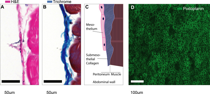
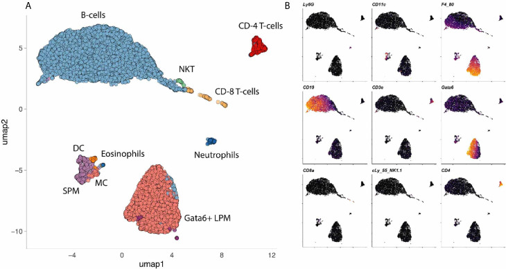
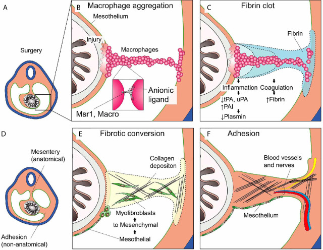
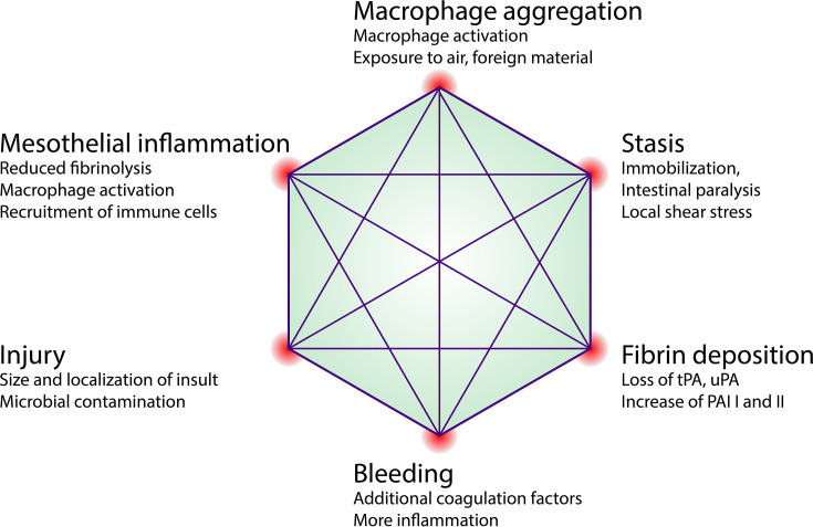
Most multicellular organisms have a major body cavity containing vital organs. This cavity is lined by a mucosa-like serosal surface and filled with serous fluid which suspends many immune cells. Injuries affecting the major body cavity are potentially life-threatening. Here we summarize evidence that unique damage detection and repair mechanisms have evolved to ensure immediate and swift repair of injuries at serosal surfaces. Furthermore, thousands of patients undergo surgery within the abdominal and thoracic cavities each day. While these surgeries are potentially lifesaving, some patients will suffer complications due to inappropriate scar formation when wound healing at serosal surfaces defects. These scars called adhesions cause profound challenges for health care systems and patients. Therefore, reviewing the mechanisms of wound repair at serosal surfaces is of clinical importance. Serosal surfaces will be introduced with a short embryological and microanatomical perspective followed by a discussion of the mechanisms of damage recognition and initiation of sterile inflammation at serosal surfaces. Distinct immune cells populations are free floating within the coelomic (peritoneal) cavity and contribute towards damage recognition and initiation of wound repair. We will highlight the emerging role of resident cavity GATA6+ macrophages in repairing serosal injuries and compare serosal (mesothelial) injuries with injuries to the blood vessel walls. This allows to draw some parallels such as the critical role of the mesothelium in regulating fibrin deposition and how peritoneal macrophages can aggregate in a platelet-like fashion in response to sterile injury. Then, we discuss how serosal wound healing can go wrong, causing adhesions. The current pathogenetic understanding of and potential future therapeutic avenues against adhesions are discussed.
Keywords: mesothelium; peritoneal adhesions; peritoneum; post-surgical adhesions; sterile injury.
Copyright © 2021 Zwicky, Stroka and Zindel.
Conflict of interest statement
JZ is holding patent rights (US63/125,020) for the use of scavenger receptor inhibitors to treat post-surgical peritoneal adhesions. The remaining authors declare that the research was conducted in the absence of any commercial or financial relationships that could be construed as a potential conflict of interest.
Figures

References
-
- Pansky B. Review of Medical Embryology. New York, NY: Macmillan; (1982).
-
- Abrahams AC, Dendooven A, van der Veer JW, Wientjes R, Toorop RJ, Bleys RLAW, et al. Direct Comparison of the Thickness of the Parietal Peritoneum Using Peritoneal Biopsy and Ultrasonography of the Abdominal Wall in Patients Treated With Peritoneal Dialysis. Perit Dialysis Int (2019) 39(5):455–64. 10.3747/pdi.2018.00108 - DOI - PubMed
-
- Hollenstein M, Jabareen M, Breitenstein S, Riener M-O, Clavien P-A, Bajka M, et al. Intraoperative Mechanical Characterization of Human Liver. PAMM (2009) 9(1):83–6. 10.1002/pamm.200910022 - DOI
Publication types
MeSH terms
Substances
LinkOut - more resources
Full Text Sources
Other Literature Sources
Medical