. 2021 May 13:12:667739.
doi: 10.3389/fpls.2021.667739. eCollection 2021.

GhGPAT12/ 25 Are Essential for the Formation of Anther Cuticle and Pollen Exine in Cotton (Gossypium hirsutum L.)

Affiliations

GhGPAT12/ 25 Are Essential for the Formation of Anther Cuticle and Pollen Exine in Cotton (Gossypium hirsutum L.)

Meng Zhang et al. Front Plant Sci. .

Abstract

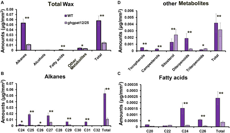
Glycerol-3-phosphate acyltransferases (GPATs), critical for multiple biological processes like male fertility, have been extensively characterized. However, their precise functions and underlying regulatory mechanism in cotton anther development are unclear. This research demonstrated the importance of GhGPAT12/25 (a paralogs pair on A12/D12 sub-chromosome of cotton) to regulate the degradation of tapetum, anther cuticle formation, and pollen exine development. GhGPAT12 and GhGPAT25 exhibited specifically detected transcripts in tapetum and pollen exine during the early anther developmental stages. GhGPAT12/25 are sn-2 glycerol-3-phosphate acyltransferases and can transfer the acyl group of palmitoyl-CoA to glycerol-3-phosphate (G3P). CRISPR/Cas9-mediated knockout identified the functional redundancy of GhGPAT12 and GhGPAT25. Knockout of both genes caused completely male sterility associated with abnormal anther cuticle, swollen tapetum, and inviable microspores with defective exine and irregular unrestricted shape. RNA-seq analysis showed that the loss of function of GhGPAT12/25 affects the processes of wax metabolic, glycerol monomer biosynthesis, and transport. Consistently, cuticular waxes were dramatically reduced in mutant anthers. Yeast one-hybrid system (Y1H), virus-induced gene silencing (VIGS), and dual-luciferase (LUC) assays illustrated that GhMYB80s are likely to directly activate the expression of GhGPAT12/25. This study provides important insights for revealing the regulatory mechanism underlying anther development in cotton.

Keywords: CRISPR/Cas9; GhGPAT12/25; anther cuticle; cotton; male sterility; pollen exine.


Conflict of interest statement

The authors declare that the research was conducted in the absence of any commercial or financial relationships that could be construed as a potential conflict of interest.

Figures

FIGURE 1
FIGURE 1
Expression pattern of GhGPAT12/25. (A) Expression levels of GhGPAT12/25 based on RNA-seq data. Error bars indicate ± S.D. (n = 3 biological replicates). (B) qRT-PCR analysis of GhGPAT12/25 in different developmental stage anthers and other tissues. Error bars indicate ± S.D. (n = 3 biological replicates). (C–G) RNA in situ hybridization assay of GhGPAT12/25 in WT anthers of panel (C) microspore mother cell (MMC) stage, (D) meiosis cell (MC) stage, (E) tetrad pollen (TTP) stage, (F) uninucleate pollen (UNP) stage, and (G) binucleate pollen (BNP) stage. (H) Anther of uninucleate pollen (UNP) stage hybridized with sense probe. MMC, microspore mother cell; MC, meiosis cell; Msp, microspore; T, tapetum; Tds, tetrads. The red arrows show positive signal in anthers. Bar = 50 μm.
FIGURE 2
FIGURE 2
Gene and protein characterization of GhGPAT12/25. (A) Structure of GhGPAT12/25 genes. Black boxes and lines indicate exons and introns, respectively. (B) Phylogenetic tree analysis of GhGPAT12/25 and their homologous sequences from other species. (C) Multiple sequence alignment of GPAT amino acids from various species. DXDX[T/V][L/V] and K-[G/S][D/S]XXX[D/N] are important motif in the haloacid dehalogenase (HAD)-like domain. AT motifs I–IV are conserved regions in acyltransferase domain. Asterisks indicate critical sites in the HAD-like domain that are indispensable for phosphatase activity of GPATs. Binding and catalytic residues in acyltransferase domain are marked by dots and triangles, respectively. (D) GPAT activity in yeast homogenates of gat-1Δ mutants transformed with either the empty pYES2 vector or the recombined plasmids containing the GhGPAT12/25 genes. Error bars indicate ± S.D. (n = 3 biological replicates).
FIGURE 3
FIGURE 3
Functional characterization of GhGPAT12/25 by CRISPR/Cas9-mediated knockout assay. (A) Gene structure of GhGPAT12/25 and the target sites of sgRNAs in exon1. The GGG and AGG in red represent the PAM motifs. (B) Variation information of transgenic lines T0-1, 7, 11, and 15. The PAM sequence is shown in red. Deletions are denoted with red dashes. Insertions are shown as red letters. The mutation types are shown on the right. L InDel indicates large InDel. (C–H) Phenotypic characteristics of ghgpat12/25 and its wild-type HM-1. (C) Adult plant, (E) flower, and (G) pollen phenotypes of the wild-type HM-1; (D) adult plant, (F) flower, and (H) pollen phenotypes of ghgpat12/25.
FIGURE 4
FIGURE 4
Paraffin section of anthers in different developmental stages of ghgpat12/25 and HM-1. Transverse section images of HM-1 anthers shown in panels (A,C,E,G,I,K) and those for ghgpat12/25 anthers shown in panels (B,D,F,H,J,L). BNP, binucleate pollen; MMC, microspore mother cell; MC, meiosis cell; MP, mature pollen; Msp, microspore; T, tapetum; Tds, tetrads; TTP, tetrad pollen; UNP, uninucleate pollen. The red arrows show main differences in anthers. Bar = 100 μm.
FIGURE 5
FIGURE 5
Scanning electron microscope (SEM) analysis of the anther surfaces and pollen grains in HM-1 and ghgpat12/25. Anthers of panels (A) HM-1 and (E) ghgpat12/25 at maturation stage. The enlarged detailed view of the anther surfaces of panels (B) HM-1 and (F) ghgpat12/25. Pollen grain of panels (C) HM-1 and (G) ghgpat12/25 at maturation stage. The enlarged detailed views of the pollen surfaces of panels (D) HM-1 and (H) ghgpat12/25.
FIGURE 6
FIGURE 6
Transmission electron microscopy (TEM) analysis of anthers in HM-1 and ghgpat12/25. (A,E,K,Q) Cross sections of the HM-1 anther cell wall at the (A) TTP stage, (E) eUNP stage, (K) lUNP stage, and (Q) BNP stage. (C,H,N,T) Cross sections of the ghgpat12/25 anther cell wall at the (C) TTP stage, (H) eUNP stage, (N) lUNP stage, and (T) BNP stage. (B,D) Tds of panel (B) HM-1 and (D) ghgpat12/25. (F,L,R) Pollen grains of HM-1 at the (F) eUNP stage, (L) lUNP stage, and (R) BNP stage. (I,O,U) Pollen grains of ghgpat12/25 at the (I) eUNP stage, (O) lUNP stage, and (U) BNP stage. (G,M,S) Pollen exine of HM-1 at the (G) eUNP stage, (M) lUNP stage, and (S) BNP stage. (J,P,V) Pollen grains of ghgpat12/25 at (J) eUNP stage, (P) lUNP stage, and (V) BNP stage. Ba, bacula; Ex, exine; In, intine; Ml, middle layer; Msp, microspore; Ne, nexine; T, tapetum; Tds, tetrads; Te, tectum. The red arrows show main differences between HM-1 and ghgpat12/25 in tapetum, tetrads, pollen grains and exine. Bars: 10 μm in panels (A-C,F,I) and (L-N,Q,T), 20 μm in panels (D,G,J) and (O,R,U), and 2 μm in panels (E,H,K,P,S,V).
FIGURE 7
FIGURE 7
TUNEL assay for the detection of DNA fragmentation in anthers of HM-1 and ghgpat12/25. (A–E) DNA fragmentation in HM-1 anthers at the (A) meiosis cell (MC) stage, (B) tetrad pollen (TTP) stage, (C) uninucleate pollen (UNP) stage, (D) binucleate pollen (BNP) stage, and (E) mature pollen (MP) stage. (F–J) DNA fragmentation in ghgpat12/25 anthers at the (F) meiosis cell (MC) stage, (G) tetrad pollen (TTP) stage, (H) uninucleate pollen (UNP) stage, (I) binucleate pollen (BNP) stage, and (J) mature pollen (MP) stage. Msp, microspore; T, tapetum; Tds, tetrads. The white arrows show positive signal in anthers. Bar = 20 μm.
FIGURE 8
FIGURE 8
RNA-seq and qRT-PCR analysis of anthers in HM-1 and ghgpat12/25. (A) Gene Ontology (GO) analysis of down-regulated and up-regulated DEGs. (B) qRT-PCR analysis of DEGs related to the synthesis and transport of lipidic monomers required for cuticle formation. (C) Biological process enrichment analysis of PPI DEGs by ClueGO plug-in in Cytoscape. The size of the nodes represents the number of genes. Node color, from blue to red, indicates increase in significance of biological processes. Error bars indicate ± S.D. (n = 3 biological replicates).
FIGURE 9
FIGURE 9
Analysis of anther waxes compositions in the anthers of WT and ghgpat12/25. (A) Total wax, (B) alkanes, (C) fatty acids, and (D) other metabolites’ amounts per unit surface area (μg/mm2) in WT and ghgpat12/25 anthers. Student’s t-test, *P < 0.05 and **P < 0.01. Error bars indicate ± S.D. (n = 3 biological replicates).
FIGURE 10
FIGURE 10
Regulatory relationship between GhGPAT12/25 and GhMYB80. (A) Transcription factors showing similar expression pattern with GhGPAT12/25. (B) Binding site sequences of MYB and bHLH (MYC) TFs on the promoters of GhGPAT12/25. The binding sites of MYB and bHLH (MYC) are located on the complementary strands. (C) Interaction between upstream TFs and the probably targeting promoter sequences of GhGPAT12/25 in yeast one-hybrid assay. (D) Expression pattern of GhMYB80s. (E) The proportion of normal anthers in pCLCrVA and pCLCrVA-GhMYB80s lines. (F) Flower phenotype of pCLCrVA and pCLCrVA-GhMYB80s lines. The red arrows show normal anthers. (G) Relative expression levels of GhMYB80s in pCLCrVA and pCLCrVA-GhMYB80s lines. (H) Relative expression levels of GhGPAT12/25 in pCLCrVA and pCLCrVA-GhMYB80s lines. (I) The relative LUC/REN ratios of the control and test groups. In panels (D,E,G,H), error bars indicate ± S.D. (n = 3 biological replicates). In panel (I), the data are the mean ± S.D. of six independent biological replicates. Student’s t-test, **P < 0.01.

Similar articles

Cited by

References

    1. Aarts M. G., Keijzer C. J., Stiekema W. J., Pereira A. (1995). Molecular characterization of the CER1 gene of Arabidopsis involved in epicuticular wax biosynthesis and pollen fertility. Plant Cell 7 2115–2127. 10.1105/tpc.7.12.2115 - DOI - PMC - PubMed
    1. Ariizumi T., Hatakeyama K., Hinata K., Sato S., Kato T., Tabata S., et al. (2003). A novel male-sterile mutant of Arabidopsis thaliana, faceless pollen-1, produces pollen with a smooth surface and an acetolysis-sensitive exine. Plant Mol. Biol. 53 107–116. 10.1023/b:plan.0000009269.97773.70 - DOI - PubMed
    1. Broun P., Poindexter P., Osborne E., Jiang C. Z., Riechmann J. L. (2004). WIN1, a transcriptional activator of epidermal wax accumulation in Arabidopsis. Proc. Natl. Acad. Sci. U S A. 101 4706–4711. 10.1073/pnas.0305574101 - DOI - PMC - PubMed
    1. Chen W., Yu X. H., Zhang K., Shi J., De Oliveira S., Schreiber L., et al. (2011). Male Sterile2 encodes a plastid-localized fatty acyl carrier protein reductase required for pollen exine development in Arabidopsis. Plant Physiol. 157 842–853. 10.1104/pp.111.181693 - DOI - PMC - PubMed
    1. Chen X., Chen G., Truksa M., Snyder C. L., Shah S., Weselake R. J. (2014). Glycerol-3-phosphate acyltransferase 4 is essential for the normal development of reproductive organs and the embryo in Brassica napus. J. Exp. Bot. 65 4201–4215. 10.1093/jxb/eru199 - DOI - PMC - PubMed