Comparative Study
. 2021 May 16:2021:3772129.
doi: 10.1155/2021/3772129. eCollection 2021.

Automatic Segmentation of Left Ventricle in Echocardiography Based on YOLOv3 Model to Achieve Constraint and Positioning

Affiliations
Comparative Study

Automatic Segmentation of Left Ventricle in Echocardiography Based on YOLOv3 Model to Achieve Constraint and Positioning

Zhemin Zhuang et al. Comput Math Methods Med. .

Abstract

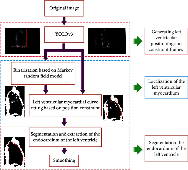
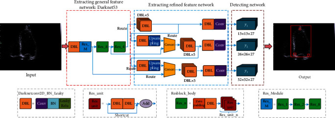
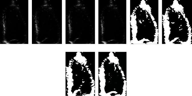
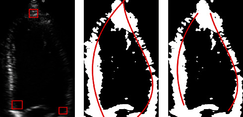
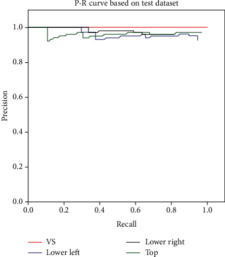
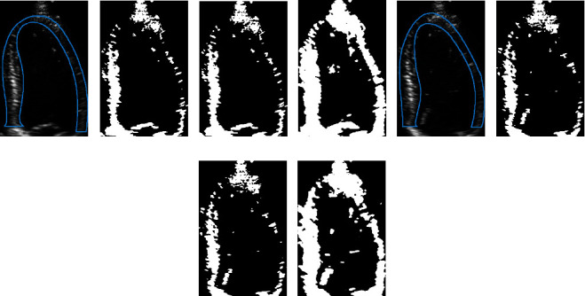
Cardiovascular disease (CVD) is the most common type of disease and has a high fatality rate in humans. Early diagnosis is critical for the prognosis of CVD. Before using myocardial tissue strain, strain rate, and other indicators to evaluate and analyze cardiac function, accurate segmentation of the left ventricle (LV) endocardium is vital for ensuring the accuracy of subsequent diagnosis. For accurate segmentation of the LV endocardium, this paper proposes the extraction of the LV region features based on the YOLOv3 model to locate the positions of the apex and bottom of the LV, as well as that of the LV region; thereafter, the subimages of the LV can be obtained, and based on the Markov random field (MRF) model, preliminary identification and binarization of the myocardium of the LV subimages can be realized. Finally, under the constraints of the three aforementioned positions of the LV, precise segmentation and extraction of the LV endocardium can be achieved using nonlinear least-squares curve fitting and edge approximation. The experiments show that the proposed segmentation evaluation indices of the method, including computation speed (fps), Dice, mean absolute distance (MAD), and Hausdorff distance (HD), can reach 2.1-2.25 fps, 93.57 ± 1.97%, 2.57 ± 0.89 mm, and 6.68 ± 1.78 mm, respectively. This indicates that the suggested method has better segmentation accuracy and robustness than existing techniques.


Conflict of interest statement

The authors declare that there are no conflicts of interest regarding the publication of this paper.

Figures

Figure 1
Figure 1
Block diagram of the proposed method.
Figure 2
Figure 2
Detection of LV based on YOLOv3.
Figure 3
Figure 3
Binarization results of LV endocardium images based on MRF: (a–d) the original frames extracted at the equal interval from the same echocardiogram and (e–h) the corresponding binarization results.
Figure 4
Figure 4
Fitting curve results of the LV subgraph in different sequences without and with constraints.
Figure 5
Figure 5
Extraction and segmentation of LV endocardium. (a) Result of binarization of the LV image using MRF model. (b) Morphological mask generated according to the fitted positioning curve. (c) Binary myocardial image obtained after mask processing. (d) Approximation result of the endocardium based on the three constraint points. (e) Result of smoothing the myocardium based on the B-spline method.
Figure 6
Figure 6
PR curve of the LV identification and constraint box based on the proposed method in this paper. (VS: ventriculus sinister; Lower left: the constraint point in the lower left corner; Lower right: the constraint point in the lower right corner; and Top: the constraint point on the top of the myocardial wall).
Figure 7
Figure 7
Binarization results of ultrasonic LV images using the Otsu method, K-means clustering method, and MRF model. The area enclosed by the blue line in (a) and (e) is the gold standard for the LV myocardium. (b, f) The binarization results obtained by using the Otsu method. (c, g) The binarization results obtained by using the K-means clustering algorithm. (d, h) The binarization results obtained by using the method proposed in this paper.

References

    1. Mensah G. A., Roth G. A., Fuster V. The global burden of cardiovascular diseases and risk factors. Journal of the American College of Cardiology. 2019;74(20):2529–2532. doi: 10.1016/j.jacc.2019.10.009. - DOI - PubMed
    1. Benyounes N., Van Der Vynckt C., Tibi S., et al. Echocardiography in confirmed and highly suspected symptomatic COVID-19 patients and its impact on treatment change. Cardiology research and practice. 2020;2020:9.4348598 - PMC - PubMed
    1. Tirronen V., Neri F., Krkkinen T., Majava K., Rossi T. An enhanced memetic differential evolution in filter design for defect detection in paper production. Evolutionary Computation. 2008;16(4):529–555. doi: 10.1162/evco.2008.16.4.529. - DOI - PubMed
    1. Caponio A., Neri F., Tirronen V. Super-fit control adaptation in memetic differential evolution frameworks. Soft Computing. 2009;13(8):811–831.
    1. Ng P. C., Henikoff S. SIFT: predicting amino acid changes that affect protein function. Nucleic Acids Research. 2003;31(13):3812–3814. doi: 10.1093/nar/gkg509. - DOI - PMC - PubMed

MeSH terms

LinkOut - more resources