

Characterization and Optimization of Benzimidazopyrimidine and Pyridoimidazopyridine Derivatives as Tau-SPECT Probes
- PMID: 34055229
- PMCID: PMC8155298
- DOI: 10.1021/acsmedchemlett.1c00071
Characterization and Optimization of Benzimidazopyrimidine and Pyridoimidazopyridine Derivatives as Tau-SPECT Probes
Abstract
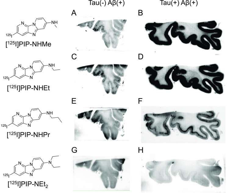
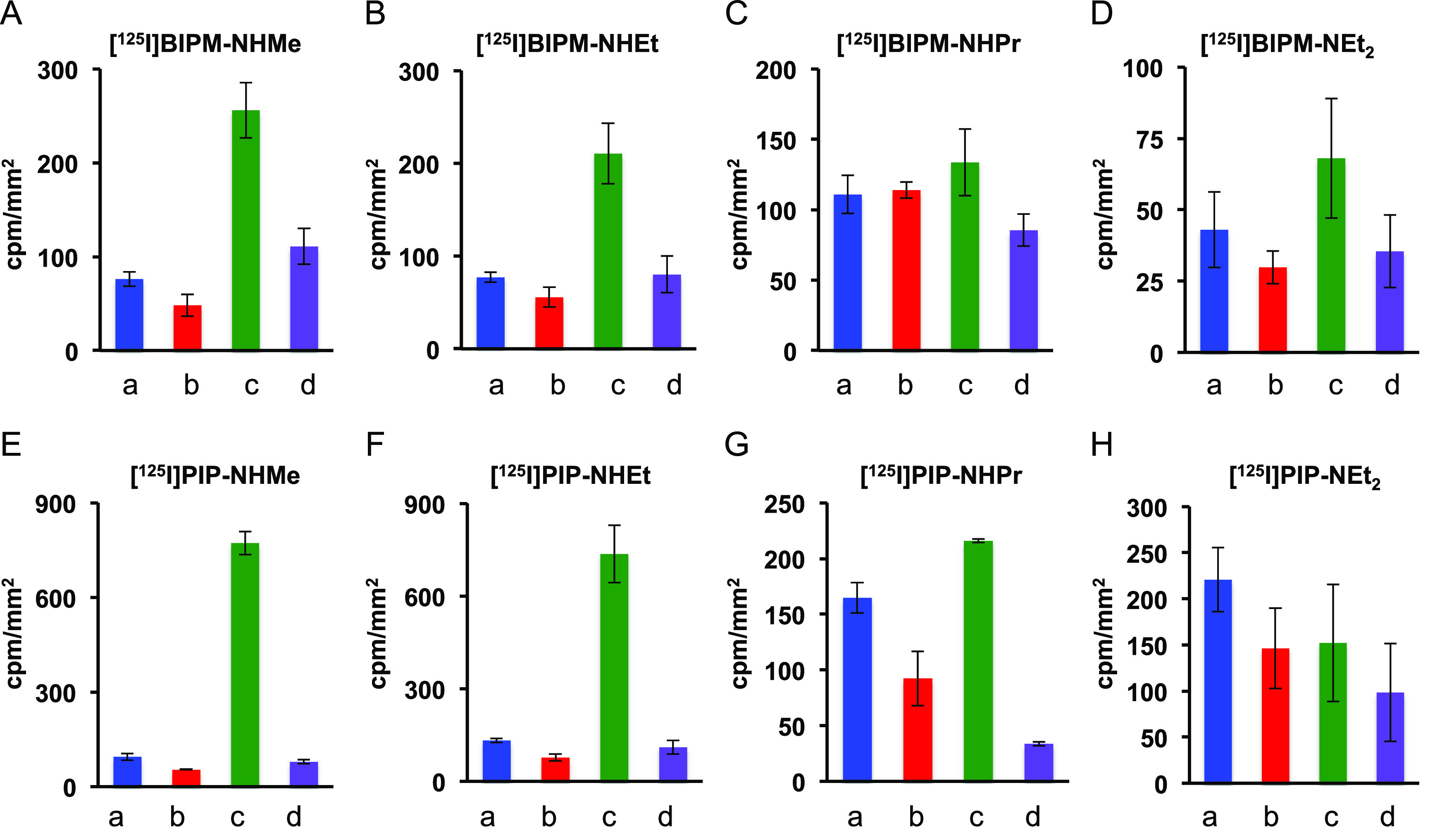
The accumulation of hyperphosphorylated tau protein in the brain is regarded as one of the hallmarks of Alzheimer's disease (AD). In vivo imaging of tau aggregates is helpful for diagnosis and monitoring of the progression of AD. In this study, we designed and synthesized novel radioiodinated benzimidazopyrimidine (BIPM) and pyridoimidazopyridine (PIP) derivatives with a monomethylamino, monoethylamino, monopropylamino, or diethylamino group as tau imaging probes for single-photon-emission computed tomography (SPECT). On in vitro autoradiography with AD brain sections, [125I]PIP-NHMe showed the highest selective binding affinity for tau aggregates among the radioiodinated BIPM and PIP derivatives. In a biodistribution study using normal mice, [125I]PIP-NHMe and [125I]PIP-NHEt displayed high initial uptake (6.62 and 6.86% ID/g, respectively, at 2 min postinjection) into and rapid clearance from the brain, with brain2 min/brain30 min ratios of 38.9 and 28.6, respectively. These results suggest that [123I]PIP-NHMe may be a novel SPECT probe that is useful for detecting tau aggregates in the AD brain.
© 2021 American Chemical Society.
Conflict of interest statement
The authors declare no competing financial interest.
Figures
Similar articles
-
Structure-Activity Relationships of Radioiodinated 6,5,6-Tricyclic Compounds for the Development of Tau Imaging Probes.ACS Med Chem Lett. 2020 Jan 9;11(2):120-126. doi: 10.1021/acsmedchemlett.9b00456. eCollection 2020 Feb 13. ACS Med Chem Lett. 2020. PMID: 32071677 Free PMC article.
-
Highly Selective Tau-SPECT Imaging Probes for Detection of Neurofibrillary Tangles in Alzheimer's Disease.Sci Rep. 2016 Sep 30;6:34197. doi: 10.1038/srep34197. Sci Rep. 2016. PMID: 27687137 Free PMC article.
-
Structure-Activity Relationships of Radioiodinated Benzoimidazopyridine Derivatives for Detection of Tau Pathology.ACS Med Chem Lett. 2018 Apr 10;9(5):478-483. doi: 10.1021/acsmedchemlett.8b00092. eCollection 2018 May 10. ACS Med Chem Lett. 2018. PMID: 29795763 Free PMC article.
-
[Development of SPECT Probes for In Vivo Imaging of β-Amyloid and Tau Aggregates in the Alzheimer's Disease Brain].Yakugaku Zasshi. 2017;137(11):1361-1365. doi: 10.1248/yakushi.17-00156. Yakugaku Zasshi. 2017. PMID: 29093372 Review. Japanese.
-
SPECT and PET imaging in Alzheimer's disease.Ann Nucl Med. 2018 Nov;32(9):583-593. doi: 10.1007/s12149-018-1292-6. Epub 2018 Aug 20. Ann Nucl Med. 2018. PMID: 30128693 Review.
Cited by
-
Recent Developments in Positron Emission Tomography Tracers for Proteinopathies Imaging in Dementia.Front Aging Neurosci. 2022 Jan 3;13:751897. doi: 10.3389/fnagi.2021.751897. eCollection 2021. Front Aging Neurosci. 2022. PMID: 35046791 Free PMC article. Review.
References
-
- Prince M.; Wimo A.; Guerchet M.; Ali G.-C.; Wu Y.-T.; Prina M., World Alzheimer Report 2015: The Global Impact of Dementia: An Analysis of Prevalence, Incidence, Cost and Trends; Alzheimer’s Disease International: London, 2015.
LinkOut - more resources
Full Text Sources
Other Literature Sources
