Review
. 2021 Apr 16;11(3):20200057.
doi: 10.1098/rsfs.2020.0057. eCollection 2021 Jun 6.

Tissue tectonics and the multi-scale regulation of developmental timing

Affiliations
Review

Tissue tectonics and the multi-scale regulation of developmental timing

Lara Busby et al. Interface Focus. .

Abstract

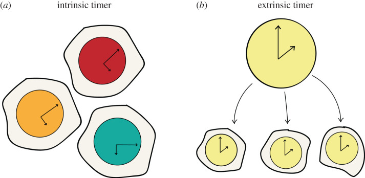
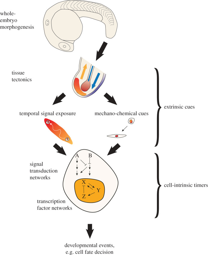
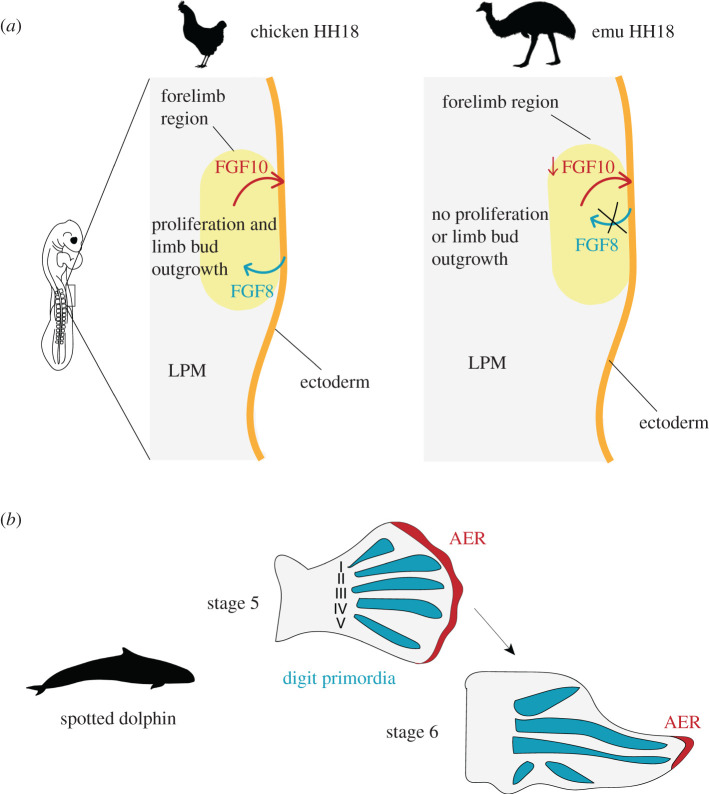
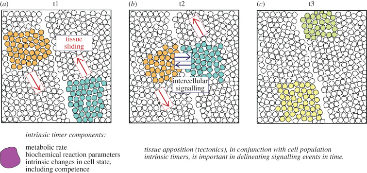
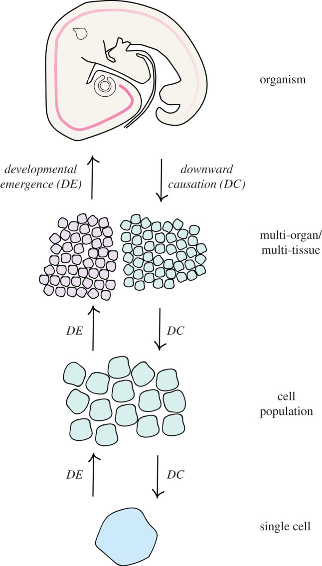
Development encompasses processes that occur at multiple length scales, including gene-regulatory interactions, cell movements and reorganization, cell signalling and growth. It is essential that the timing of events in all of these different processes is coordinated to generate well-patterned tissues and organs. However, how the timing of intrinsic cell state changes is coordinated with events occurring at the multi-tissue and whole-organism level is unknown. Here, we argue that an important mechanism that accounts for the integration of timing across levels of organization is provided by tissue tectonics, i.e. how morphogenetic events driving tissue shape changes result in the relative displacement of signalling and responding tissues and coordinate developmental timing across scales. In doing so, tissue tectonics provides a mechanism by which the cell specification events intrinsic to cells can be modulated by the temporal exposure to extracellular signals. This exposure is in turn regulated by higher-order properties of the embryo, such as their physical properties, rates of growth and the combination of dynamic cell behaviours, impacting tissue morphogenesis. Tissue tectonics creates a downward flow of information from higher to lower levels of biological organization, providing an instance of downward causation in development.

Keywords: developmental; multi-scale; review; tectonics; timings; tissue.


Figures

Figure 1.
Figure 1.
Intrinsic and extrinsic timers. Schematic summarizing the distinction between intrinsic and extrinsic timers. Each of the ‘blobs’ represents either a cell population or cell, dependent upon context. In an intrinsic timer mechanism, each population (cell) has its own internal timer, which is not affected by external information. By contrast, in an extrinsic timer mechanism, information from the surroundings is integrated by the population (cell) to infer the state of the timer.
Figure 2.
Figure 2.
Mechanisms of downward causation in development. This schematic shows the different ways in which morphogenesis can influence cell-intrinsic transcription factor networks and ultimately the timing of developmental events. This can occur both via mechano-chemical cues (as described elsewhere—see text for references), and, as we propose here, through the modulation of temporal signal exposure. As whole-embryo morphogenesis occurs (top panel), signalling and responding tissues move relative to one another (tissue tectonics, second panel). Thus, the exposure of the responding tissue to signals changes over time. Signals feed into cell-intrinsic signal transduction networks that ultimately converge on transcription factor networks. In this way, extrinsic cues interface with cell-intrinsic timing mechanisms via tissue tectonics.
Figure 3.
Figure 3.
Heterochrony between tissues can produce new forms in evolution. (a) Schematic of the chicken and emu lateral plate mesoderm (LPM) at HH18. In most avian species, including the chicken, reciprocal signalling between the forelimb region of the LPM and the overlying ectoderm (orange) promotes proliferation of LPM cells and limb bud outgrowth. However, the emu forelimb region of the LPM produces the signalling ligand FGF10 at an insufficient level to induce FGF8 production in the ectoderm (orange). Consequently, cells of the LPM forelimb region do not proliferate and the limb bud does not grow out from the body at HH17. (b) Analysis of spotted dolphin developing limb buds has shown that the apical ectodermal ridge (AER) persists over digits II and III for longer than over the other digits. These digits exhibit hyperphalangy (many finger bones) and it has been suggested that the local persistence of the AER may be related to this trait. Silhouette images of animals were taken from PhyloPic: please see Acknowledgements for attributions.
Figure 4.
Figure 4.
Tissue tectonics. Three snapshots in time are shown in this schematic, at time points t1, t2 and t3. Two populations of cells are shown in orange and teal, each of which is found in different tissue sheets. As the tissues slide relative to one another, the populations of interest come into close apposition, allowing intercellular signalling to occur. At t3, the inductive signalling event has occurred and both populations' cells have experienced a change in cell state. Clearly, tissue tectonics is an important contributor to the timing of signalling events in development.
Figure 5.
Figure 5.
Summary. This schematic summarizes the different levels of organization where timing has been studied in developmental biology. Interaction between the various levels of organization occurs bidirectionally. Information passes from higher to lower levels through downward causation, exemplified by pattern regulation in the embryo (see text for examples). Information also passes from the lower levels to higher levels, through developmental emergence. For example, changes in cell state can lead to changes to inter-population signalling and ultimately direct higher-level changes to the embryo in morphogenesis. We argue that a link between these levels of organization is provided by the concept of tissue tectonics. The ways in which signalling and responding tissues are displaced relative to one another can influence the timing and location of signalling events between tissues.

References

    1. Duboule D. 2003. Time for chronomics? Science 301, 277. (10.1126/science.301.5631.277) - DOI - PubMed
    1. Ebisuya M, Briscoe J. 2018. What does time mean in development? Development 145, dev164368. (10.1242/dev.164368) - DOI - PMC - PubMed
    1. Johnson MH, Day ML. 2000. Egg timers: how is developmental time measured in the early vertebrate embryo? Bioessays 22, 57-63. (10.1002/(SICI)1521-1878(200001)22:1<57::AID-BIES10>3.0.CO;2-L) - DOI - PubMed
    1. Slack J. 1983. From egg to embryo. Cambridge, UK: Cambridge University Press.
    1. Linker C, Stern CD. 2004. Neural induction requires BMP inhibition only as a late step, and involves signals other than FGF and Wnt antagonists. Development 131, 5671-5681. (10.1242/dev.01445) - DOI - PubMed

LinkOut - more resources