Review
. 2020 Nov 29;8(3):247-258.
doi: 10.1093/nop/npaa079. eCollection 2021 Jun.

Case series review of neuroradiologic changes associated with immune checkpoint inhibitor therapy

Affiliations
Review

Case series review of neuroradiologic changes associated with immune checkpoint inhibitor therapy

Na Tosha N Gatson et al. Neurooncol Pract. .

Abstract

While immuno-oncotherapy (IO) has significantly improved outcomes in the treatment of systemic cancers, various neurological complications have accompanied these therapies. Treatment with immune checkpoint inhibitors (ICIs) risks multi-organ autoimmune inflammatory responses with gastrointestinal, dermatologic, and endocrine complications being the most common types of complications. Despite some evidence that these therapies are effective to treat central nervous system (CNS) tumors, there are a significant range of related neurological side effects due to ICIs. Neuroradiologic changes associated with ICIs are commonly misdiagnosed as progression and might limit treatment or otherwise impact patient care. Here, we provide a radiologic case series review restricted to neurological complications attributed to ICIs, anti-CTLA-4, and PD-L-1/PD-1 inhibitors. We report the first case series dedicated to the review of CNS/PNS radiologic changes secondary to ICI therapy in cancer patients. We provide a brief case synopsis with neuroimaging followed by an annotated review of the literature relevant to each case. We present a series of neuroradiological findings including nonspecific parenchymal and encephalitic, hypophyseal, neural (cranial and peripheral), meningeal, cavity-associated, and cranial osseous changes seen in association with the use of ICIs. Misdiagnosis of radiologic abnormalities secondary to neurological immune-related adverse events can impact patient treatment regimens and clinical outcomes. Rapid recognition of various neuroradiologic changes associated with ICI therapy can improve patient tolerance and adherence to cancer therapies.

Keywords: autoimmune encephalitis; brain metastases; immune checkpoint inhibitors (ICIs); immune-related adverse events (irAEs); neuroradiology.


Figures

Figure 1.
Figure 1.
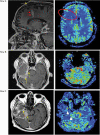
Case of nonspecific asymptomatic changes (A-C). (A) Within the first month of ICI therapy. Sagittal view of T1 FLAIR propeller post-contrast demonstrating normal brain. (B) Within 3 months of initiating ICI therapy. Sagittal view of T1 3D SPGR post-contrast demonstrating the appearance of left cerebellar lesion (yellow circle). The initial radiographic impression read, “potentially immunotherapy-related treatment-related, however, metastatic disease cannot be ruled out.” (C) Eight months after discontinuing ICI therapy. Sagittal view of T1 3D SPGR post-contrast demonstrating resolution of the previously noted enhancing left cerebellar lesion. Case of peri-tumoral changes (rows D-G). (Row D) 6 months after starting ICI. Interval size increase of the lesion in the left parieto-occipital junction (Row D, left). The edema surrounding this lesion has also increased yellow circle. A 5-mm enhancing lesion (T1 post-contrast not shown) with associated peritumoral edema in the right occipital lobe appears slightly smaller than on prior study (top right, red oval). (Row E) Also at 6 months after the start of ICI—correlate perfusion-weighted imaging does not demonstrate elevation in rCBV associated with either the bilateral occipital lobe areas yellow circle left and red oval on right. More consistent with a treatment-related effect. (Row F) 14 months after starting ICI. The left parieto-occipital lesion has slightly decreased in size, with less surrounding T2/FLAIR signal abnormality (middle left). Slight interval increase in the size of the right occipital lobe peritumoral edema without changes in T1 contrast (not shown) (middle right). (Row G) 19 months’ follow-up after starting ICI with noted further decrease in size of T2 FLAIR signal abnormality in the left temporal occipital region (bottom left). Further interval fluctuation in size and prominence of right occipital lobe FLAIR abnormality representing nonspecific peritumoral vasogenic edema (bottom right).
Figure 2.
Figure 2.
Case of peri-device and peri-resection cavity changes. MRI brain left-side T1 post-contrast and right-side perfusion-weighted imaging. (Row A) Peri-device changes (left) sagittal image Ommaya reservoir (yellow arrow) with enhancement along catheter (red arrows), without elevated rCBV (red oval) (right), described as possible disseminating necrotizing leukoencephalopathy (DNL) vs peri-device associated inflammation secondary to ICI, no concern for malignancy. (Row B) Peri-resection cavity changes (left) axial image with diffuse linear enhancement along the cavity (yellow arrows) without nodularity or associated elevated rCBV (right). (Row C) (left) axial image with continued progression of enhancement with increased nodularity (yellow arrows) around resection cavity and mild elevation of rCBV (white arrowheads) on perfusion study (right)—likely combined treatment-related encephalitis and tumor progression.
Figure 3.
Figure 3.
Case of metastatic mimicry. Preoperative MRI 1 year after starting anti-PD-1 immunotherapy. (A) Axial T1 post-contrast, (B) T2 FLAIR, (C) T2 fat saturation demonstrate a septated “mass” lesion with heterogeneous enhancement and lobulated contour centered within the right occipital lobe with surrounding vasogenic edema and mild local mass effect. (D) Diffuse perivascular infiltrate of small-sized monotonous lymphocytes with mature chromatin (scale bar 100 µm). (E) CD4-positive staining of T cells (scale bar 100 µm).
Figure 4.
Figure 4.
Case of hypophysitis. Sagittal T1 post-contrast brain MRI. (A) One week prior to starting anti-CTLA-4 therapy demonstrating the absence of acute intracranial abnormalities. (B) Two months after initiation of therapy demonstrating enlargement of the pituitary gland (yellow arrow). (C) Two months after discontinuation of ICIs demonstrating interval resolution of pituitary enlargement.
Figure 5.
Figure 5.
Case of optic neuritis and nonspecific changes (A-G). MRI T1 thin FAT SAT plus contrast (A) coronal and (B) axial orbits with homogeneous enhancement of the intracranial segments of the optic nerves (yellow arrows). (C) and (D) nonspecific enhancing lesions of the cerebellum. (E-G) Follow-up imaging (2.5 months later) demonstrated interval resolution of nonspecific imaging changes along the optic nerves (E, red circle) and in the cerebellum (G, yellow circle). However, new findings on follow-up identified a new enhancing lesion in the right temporal lobe with associated elevation in rCBV (E/F yellow arrows) and vasogenic edema on T2 sequences (not shown). Case of brachial neuritis (Parsonage-Turner Syndrome) (H, I). (H) Coronal T2-weighted images with fat saturation show increased T2 signal in the supraspinatus, infraspinatus, and teres minor (red arrowheads) muscles consistent with edema. MRI chest, brachial plexus (I) axial STIR with diffusely increased signal in the left infraspinatus muscle (yellow rectangle) consistent with denervation from brachial neuritis.
Figure 6.
Figure 6.
Case of vascular malformation and spinal cord expansion. MRI sagittal STIR (yellow arrows) 6 months after initiation of ICI (A), T2 (B), and axial T2 (C) with noted diffusely increased signal within the mildly enlarged distal spinal cord primarily in the region of the conus with noted primary central cord edema. Findings are nonspecific and differential includes inflammation, demyelination, infection, or neoplastic etiologies. MRA/MRI T/L spine 3 months symptomatic. (D) Sagittal T2 image with continued mild distal expansion and dilated perimedullary vessels (yellow arrows) (E) sagittal and (F) axial T1 post-contrast with intense contrast enhancement of the distal spinal cord and conus medullaris (red arrows), and (G) enhancement of the dorsal perimedullary veins (thick yellow arrows). Findings are characteristic of a spinal dAVF (type 1 AVM).
Figure 7.
Figure 7.
Case of pachymeningitis (rows A-D). (Row A) Axial T1 post-contrast MRI brain 3 months prior to re-initiation of therapy. (Row B) Axial T1 post-contrast MRI brain 6 months after re-initiation of ICI demonstrating multifocal leptomeningeal and dural signal abnormalities and enhancement. (Row C) Contrasted MRI brain axial (left) perfusion-weighted imaging without elevation in rCBV in corresponding areas of worsening enhancement (right). (Row D) Axial T1 post-contrast imaging at 8-week follow-up imaging with continued progression of tumor along the meningeal and subependymal surfaces (yellow arrows) and elevated rCBV in perfusion imaging on far right. Case of osseous changes (E-G). Sagittal T1 brain MRI (E) imaging 1 month prior to start of anti-PD-1 therapy with intact bone as compared to (F) 5 months after starting ICI therapy demonstrating new diffuse T1 hypo-intensities (white arrows) of bone marrow signal in imaged osseous structures, including calvarium, skull base, and imaged upper cervical spine. (G) MRI brain follow-up 3 months off ICIs with interval partial resolution of abnormal marrow signal.

References

    1. Cohen JV, Tawbi H, Margolin KA, et al. . Melanoma central nervous system metastases: current approaches, challenges, and opportunities. Pigment Cell Melanoma Res. 2016;29(6):627–642. - PMC - PubMed
    1. Suarez-Sarmiento A Jr, Nguyen KA, Syed JS, et al. . Brain metastasis from renal-cell carcinoma: an institutional study. Clin Genitourin Cancer. 2019;17(6):e1163–e1170. - PubMed
    1. Ali A, Goffin JR, Arnold A, et al. . Survival of patients with non-small-cell lung cancer after a diagnosis of brain metastases. Curr Oncol. 2013;20(4):e300–e306. - PMC - PubMed
    1. Kotecki N, Lefranc F, Devriendt D, et al. . Therapy of breast cancer brain metastases: challenges, emerging treatments and perspectives. Ther Adv Med Oncol. 2018;10:1758835918780312. - PMC - PubMed
    1. Qin S, Xu L, Yi M, et al. . Novel immune checkpoint targets: moving beyond PD-1 and CTLA-4. Mol Cancer. 2019;18(1):155. - PMC - PubMed