

High fructose exposure modifies the amount of adipocyte-secreted microRNAs into extracellular vesicles in supernatants and plasma
- PMID: 34055478
- PMCID: PMC8140597
- DOI: 10.7717/peerj.11305
High fructose exposure modifies the amount of adipocyte-secreted microRNAs into extracellular vesicles in supernatants and plasma
Abstract
Background: High fructose exposure induces metabolic and endocrine responses in adipose tissue. Recent evidence suggests that microRNAs in extracellular vesicles are endocrine signals secreted by adipocytes. Fructose exposure on the secretion of microRNA by tissues and cells is poorly studied. Thus, the aim of this study was to evaluate the effect of fructose exposure on the secretion of selected microRNAs in extracellular vesicles from 3T3-L1 cells and plasma from Wistar rats.
Methods: 3T3-L1 cells were exposed to 550 µM of fructose or standard media for four days, microRNAs levels were determined in extracellular vesicles of supernatants and cells by RT-qPCR. Wistar rats were exposed to either 20% fructose drink or tap water for eight weeks, microRNAs levels were determined in extracellular vesicles of plasma and adipose tissue by RT-qPCR.
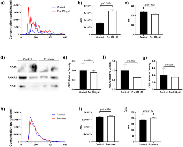
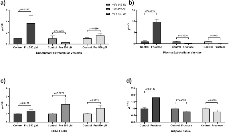
Results: This study showed that fructose exposure increased the total number of extracellular vesicles released by 3T3-L1 cells (p = 0.0001). The levels of miR-143-5p were increased in extracellular vesicles of 3T3-L1 cells exposed to fructose (p = 0.0286), whereas miR-223-3p levels were reduced (p = 0.0286). Moreover, in plasma-derived extracellular vesicles, miR-143-5p was higher in fructose-fed rats (p = 0.001), whereas miR-223-3p (p = 0.022), miR-342-3p (p = 0.0011), miR-140-5p (p = 0.0129) and miR-146b-5p (p = 0.0245) were lower.
Conclusion: Fructose exposure modifies the levels of microRNAs in extracellular vesicles in vitro and in vivo. In particular, fructose exposure increases miR-143-5p, while decreases miR-223-3p and miR-342-3p.
Keywords: Adipocytes; Adipose tissue; Extracellular Vesicles; Fructose; microRNA.
©2021 Hernández-Díazcouder et al.
Conflict of interest statement
The authors declare there are no competing interests.
Figures
Similar articles
-
Adipose mesenchymal stem cell-derived extracellular vesicles containing microRNA-26a-5p target TLR4 and protect against diabetic nephropathy.J Biol Chem. 2020 Sep 11;295(37):12868-12884. doi: 10.1074/jbc.RA120.012522. Epub 2020 Jun 24. J Biol Chem. 2020. PMID: 32580945 Free PMC article.
-
Cold exposure aggravates pulmonary arterial hypertension through increased miR-146a-5p, miR-155-5p and cytokines TNF-α, IL-1β, and IL-6.Life Sci. 2021 Dec 15;287:120091. doi: 10.1016/j.lfs.2021.120091. Epub 2021 Oct 28. Life Sci. 2021. PMID: 34717910
-
Analysis of miR-9-5p, miR-124-3p, miR-21-5p, miR-138-5p, and miR-1-3p in Glioblastoma Cell Lines and Extracellular Vesicles.Int J Mol Sci. 2020 Nov 11;21(22):8491. doi: 10.3390/ijms21228491. Int J Mol Sci. 2020. PMID: 33187334 Free PMC article.
-
miR-103a-3p and miR-22-5p Are Reliable Reference Genes in Extracellular Vesicles From Cartilage, Adipose Tissue, and Bone Marrow Cells.Front Bioeng Biotechnol. 2021 Feb 15;9:632440. doi: 10.3389/fbioe.2021.632440. eCollection 2021. Front Bioeng Biotechnol. 2021. PMID: 33659243 Free PMC article.
-
Colorectal cancer cell-derived extracellular vesicles transfer miR-221-3p to promote endothelial cell angiogenesis via targeting suppressor of cytokine signaling 3.Life Sci. 2021 Nov 15;285:119937. doi: 10.1016/j.lfs.2021.119937. Epub 2021 Sep 8. Life Sci. 2021. PMID: 34508764
Cited by
-
Diagnostic and prognostic value of miR-146b-5p in acute pancreatitis.Hereditas. 2025 May 31;162(1):93. doi: 10.1186/s41065-025-00466-9. Hereditas. 2025. PMID: 40450371 Free PMC article.
References
-
- Brianza-Padilla M, Carbó R, Arana JC, Vázquez-Palacios G, Ballinas-Verdugo MA, Cardoso-Saldaña GC, Palacio AG, Juárez-Vicuña Y, Sánchez F, Martínez-Martínez E, Huang F, Sánchez-Muñoz F, Bojalil R. Inflammation related MicroRNAs are modulated in total plasma and in extracellular vesicles from rats with chronic ingestion of sucrose. BioMed Research International. 2016;2016:1–7. doi: 10.1155/2016/2489479. - DOI - PMC - PubMed
LinkOut - more resources
Full Text Sources
Other Literature Sources
Miscellaneous
