Giant Lactating Adenoma With Fibroadenomated Changes
- PMID: 34055546
- PMCID: PMC8155741
- DOI: 10.7759/cureus.14706
Giant Lactating Adenoma With Fibroadenomated Changes
Abstract
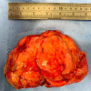
Lactating adenomas (LAs) are uncommon benign breast tumors that typically occur in the late pregnancy or lactation period and are among the most prevalent breast lesions during puerperium. They commonly present with a painless, rapidly growing, large, mobile breast lump either late in pregnancy or the postpartum period. Despite being a condition, a core biopsy is almost always required to exclude malignancy. We are presenting a case of a 34-year-old patient who was referred to our unit with a progressive increase in size of the pre-existing right breast lump that has been there before pregnancy. Due to the massive increase in size in a short period, the lump was removed shortly after delivery with an acceptable cosmetic outcome.
Keywords: benign breast disease; fibroadenomatoid change; lactating adenoma; pregnancy related breast changes.
Copyright © 2021, Monib et al.
Conflict of interest statement
The authors have declared that no competing interests exist.
Figures


References
-
- The SCARE 2018 statement: updating consensus Surgical CAse REport (SCARE) guidelines. Agha RA, Borrelli MR, Farwana R, Koshy K, Fowler AJ, Orgill DP. Int J Surg. 2018;60:132–136. - PubMed
-
- Lactating adenoma of the breast. Barco Nebreda I, Vidal MC, Fraile M, Canales L, González C, Giménez N, García-Fernández A. J Hum Lact. 2016;32:559–562. - PubMed
-
- Breast imaging of the pregnant and lactating patient: physiologic changes and common benign entities. Vashi R, Hooley R, Butler R, Geisel J, Philpotts L. Am J Roentgenol. 2013;200:329–336. - PubMed
Publication types
LinkOut - more resources
Full Text Sources
