Review
. 2021 May 13:9:676911.
doi: 10.3389/fcell.2021.676911. eCollection 2021.

Length of the Neurogenic Period-A Key Determinant for the Generation of Upper-Layer Neurons During Neocortex Development and Evolution

Affiliations
Review

Length of the Neurogenic Period-A Key Determinant for the Generation of Upper-Layer Neurons During Neocortex Development and Evolution

Barbara K Stepien et al. Front Cell Dev Biol. .

Abstract

The neocortex, a six-layer neuronal brain structure that arose during the evolution of, and is unique to, mammals, is the seat of higher order brain functions responsible for human cognitive abilities. Despite its recent evolutionary origin, it shows a striking variability in size and folding complexity even among closely related mammalian species. In most mammals, cortical neurogenesis occurs prenatally, and its length correlates with the length of gestation. The evolutionary expansion of the neocortex, notably in human, is associated with an increase in the number of neurons, particularly within its upper layers. Various mechanisms have been proposed and investigated to explain the evolutionary enlargement of the human neocortex, focussing in particular on changes pertaining to neural progenitor types and their division modes, driven in part by the emergence of human-specific genes with novel functions. These led to an amplification of the progenitor pool size, which affects the rate and timing of neuron production. In addition, in early theoretical studies, another mechanism of neocortex expansion was proposed-the lengthening of the neurogenic period. A critical role of neurogenic period length in determining neocortical neuron number was subsequently supported by mathematical modeling studies. Recently, we have provided experimental evidence in rodents directly supporting the mechanism of extending neurogenesis to specifically increase the number of upper-layer cortical neurons. Moreover, our study examined the relationship between cortical neurogenesis and gestation, linking the extension of the neurogenic period to the maternal environment. As the exact nature of factors promoting neurogenic period prolongation, as well as the generalization of this mechanism for evolutionary distinct lineages, remain elusive, the directions for future studies are outlined and discussed.

Keywords: evolution; gestation; neocortex; neurogenic period length; upper-layer neurons.


Conflict of interest statement

The authors declare that the research was conducted in the absence of any commercial or financial relationships that could be construed as a potential conflict of interest.

Figures
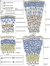
FIGURE 1
FIGURE 1
Schematic representation of the developing neocortical wall in mouse and human. (A) Mouse neocortical wall. (B) Human neocortical wall. (C) Mouse (left) and human (right) cortical plates. Note both the tangential and radial (predominantly in the upper layers) expansion of the human cortical plate. For cell types, see keys in (A,C).
FIGURE 2
FIGURE 2
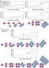
(A) Illustration of the effects of different modes of progenitor division on the number of neurons produced, showing the results after 3 cell cycles each. Left: Asymmetric neurogenic progenitor divisions (1 progenitor: >1 progenitor + 1 neuron; see diagram) lead to a linear increase in total cell number with every cell cycle (top graph, black), with the number of progenitors remaining constant (bottom graph, magenta) and the number of neurons increasing linearly after the first cell cycle (bottom graph, blue). Right: In contrast, symmetric divisions lead to an exponential increase in total cell number with every cell cycle (top graph). The initial symmetric proliferative progenitor divisions (1 progenitor: >2 progenitors; see diagram) double the number of progenitors with every cell cycle (bottom graph, magenta). Upon progenitors switching to symmetric consumptive neurogenic division (arrow), the number of neurons (bottom graph, blue) is twice that of the (now consumed) progenitors. The longer the neurogenic period and the greater the number of progenitor cell cycles, the greater the increase in the number of neurons generated in the right scenario compared to the left one. (B) Illustration of the effects of delaying the switch of symmetric proliferative progenitor divisions to asymmetric neurogenic progenitor divisions (see top and bottom diagrams). Progenitors undergo one (top) or two (bottom) cycle(s) of symmetric proliferative division and then switch to asymmetric neurogenic progenitor divisions, with the number of progenitors then generated remaining constant and the number of neurons increasing linearly. The number of neurons produced upon each asymmetric neurogenic progenitor division matches the number of progenitors present. Because the number of progenitors is twice as high after two (bottom) than one (top) cycle(s) of symmetric proliferative division, the number of neurons added upon each cycle of asymmetric neurogenic progenitor division is twice as high in the bottom than top scenario, leading to a steeper increase in neuron numbers and a greater neuron output in the bottom scenario, although neurogenesis starts one progenitor cycle later.
FIGURE 3
FIGURE 3
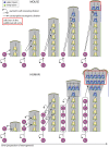
Simplified model illustrating how a selective increase in upper-layer neurons in human as compared to mouse can be achieved by changing the type of BP generated from aRG and the mode of BP division, with the same temporal progression regarding the change from deep-layer to upper-layer neuron generation. In both mouse (top) and human (bottom), aRG (bottom cells in each diagram) undergo repeated cycles of asymmetric, self-renewing and BP-genic, division. However, the type of BP generated and its mode of division over the course of neurogenesis are different. Top: With each cycle of asymmetric self-renewing division (round arrows), an aRG in embryonic mouse neocortex generates one bIP (straight vertically pointing arrows), which then undergoes consumptive neurogenic division, generating two neurons (pairs of straight oblique arrows). This leads to a linear increase in the total number of neurons generated, with two neurons added per single aRG division cycle. Bottom: With each cycle of asymmetric self-renewing division (round arrows), an aRG in fetal human neocortex generates one bRG (straight vertically pointing arrows). This undergoes repeated cycles of asymmetric, self-renewing division (round arrows), with the other daughter being a neuron (straight oblique arrows). As each bRG persists due to self-renewal, this leads to a linear increase in the number of bRG with each aRG cycle. This in turn leads to a progressive increase in the number of neurons generated per aRG cycle, which is equal to that in mouse after the 3rd aRG cycle, that is, an equal number of deep-layer neurons has then been generated in both the mouse and human scenario. However, if we assume that for both mouse and human, BPs switch to generate upper-layer neurons in the 4th aRG cycle, due to the accumulation of bRG in human, a greater number of upper-layer neurons in generated in this cycle in human than mouse. This difference becomes even greater if in the next cycle, the bRG in human, like the bIPs in mouse, adopt a consumptive neurogenic mode of division (red boxes), perhaps via the generation of bIPs (not illustrated; for a more detailed depiction of possible BP lineages, see Lewitus et al., 2014). Comments: (i) Hence, lengthening the neurogenic period, e.g., from the 4th to the 5th aRG cycle, results in a selective increase in upper-layer neurons for both mouse and human; (ii) a lineage of asymmetric self-renewing BP-genic aRG division followed by asymmetric self-renewing neurogenic bRG division followed by consumptive neurogenic bRG division (see above) results in a greater upper-layer neuron output in human than the lineage of asymmetric self-renewing BP-genic aRG division followed by consumptive neurogenic bIP division in mouse.
FIGURE 4
FIGURE 4
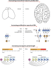
Diagram illustrating the two major factors underlying the increase, during development, in neuron production associated with the evolutionary expansion of the neocortex, as depicted for mouse vs. human in the top row (images not drawn to scale). (1) Middle row: Increase in the proliferative capacity of BPs by changing the type of BPs and their mode of division. BPs in embryonic mouse neocortex comprise mostly bIPs of which each one undergoes a single consumptive division generating two neurons (N). BPs in fetal human neocortex comprise both bRG and bIPs, both of which can undergo various modes of cell division (see Lewitus et al., 2014) of which the following are illustrated. bRG may undergo repeated asymmetric self-renewing divisions generating one neuron each. bIPs may first undergo symmetric proliferative divisions, resulting in an exponential increase in their number, followed by consumptive neurogenic divisions of these bIPs. (2) Bottom row: Selective increase in upper-layer cortical neurons upon lengthening the neurogenic period. Once progenitors (P) that generate neurons (N) have switched from generating deep-layer neurons to generating upper-layer neurons, a lengthening of the neurogenic period (red bar) during neocortex development, e.g., in fetal human as compared to embryonic mouse, will result in selectively increasing upper-layer neurons. For simplicity, and to illustrate the underlying principle, irrespective of the actual lineages in embryonic mouse vs. fetal human neocortex (see middle row), in the example illustrated, a progenitor is assumed to successively generate first deep-layer and then upper-layer neurons by repeated asymmetric self-renewing divisions, which occur for longer in fetal human than embryonic mouse neocortex. Not illustrated—for the ease of presentation—are other lineage scenarios, in which with a mixed population of progenitors, some progenitors undergo symmetric proliferative divisions while others undergo neurogenic, e.g., symmetric consumptive, divisions, with both types of progenitor divisions occurring for longer in species developing an expanded neocortex.

References

    1. Allman J. M. (1999). Evolving Brains. New York, NY: Scientifc American Library.
    1. Anderson S. A., Eisenstat D. D., Shi L., Rubenstein J. L. (1997). Interneuron migration from basal forebrain to neocortex: dependence on Dlx genes. Science 278 474–476. 10.1126/science.278.5337.474 - DOI - PubMed
    1. Angevine J. B. J., Sidman R. L. (1961). Autoradiographic study of cell migration during histogenesis of cerebral cortex in the mouse. Nature 192 766–768. 10.1038/192766b0 - DOI - PubMed
    1. Armstrong E., Curtis M., Buxhoeveden D. P., Fregoe C., Zilles K., Casanova M. F., et al. (1991). Cortical gyrification in the rhesus monkey: a test of the mechanical folding hypothesis. Cereb. Cortex 1 426–432. 10.1093/cercor/1.5.426 - DOI - PubMed
    1. Azevedo F. A. C., Carvalho L. R. B., Grinberg L. T., Farfel J. M., Ferretti R. E. L., Leite R. E. P., et al. (2009). Equal numbers of neuronal and nonneuronal cells make the human brain an isometrically scaled-up primate brain. J. Comp. Neurol. 513 532–541. 10.1002/cne.21974 - DOI - PubMed