Review
. 2021 May 14:8:659550.
doi: 10.3389/fmolb.2021.659550. eCollection 2021.

Insights into Aldehyde Dehydrogenase Enzymes: A Structural Perspective

Affiliations
Review

Insights into Aldehyde Dehydrogenase Enzymes: A Structural Perspective

Kim Shortall et al. Front Mol Biosci. .

Abstract

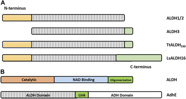
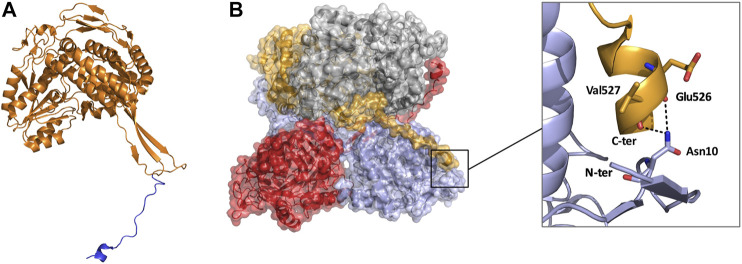
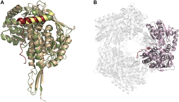
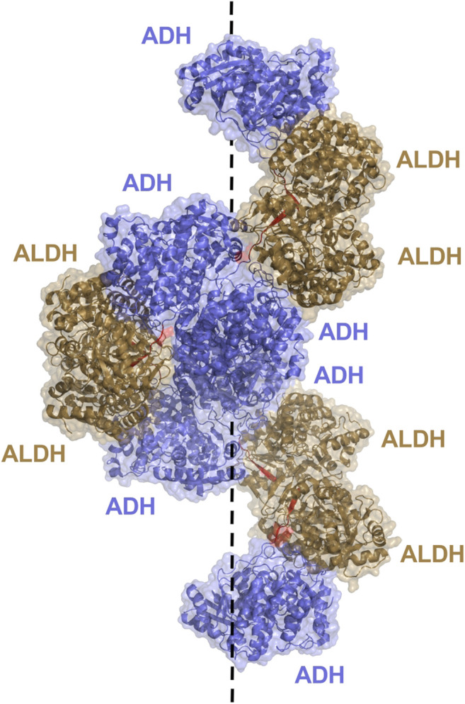
Aldehyde dehydrogenases engage in many cellular functions, however their dysfunction resulting in accumulation of their substrates can be cytotoxic. ALDHs are responsible for the NAD(P)-dependent oxidation of aldehydes to carboxylic acids, participating in detoxification, biosynthesis, antioxidant and regulatory functions. Severe diseases, including alcohol intolerance, cancer, cardiovascular and neurological diseases, were linked to dysfunctional ALDH enzymes, relating back to key enzyme structure. An in-depth understanding of the ALDH structure-function relationship and mechanism of action is key to the understanding of associated diseases. Principal structural features 1) cofactor binding domain, 2) active site and 3) oligomerization mechanism proved critical in maintaining ALDH normal activity. Emerging research based on the combination of structural, functional and biophysical studies of bacterial and eukaryotic ALDHs contributed to the appreciation of diversity within the superfamily. Herewith, we discuss these studies and provide our interpretation for a global understanding of ALDH structure and its purpose-including correct function and role in disease. Our analysis provides a synopsis of a common structure-function relationship to bridge the gap between the highly studied human ALDHs and lesser so prokaryotic models.

Keywords: C-terminal extensions; NAD(P) cofactor; aldehyde dehydrogenase; enzyme dysfunction; mutations; oligomerization; spirosomes; structure-function.


Conflict of interest statement

The authors declare that the research was conducted in the absence of any commercial or financial relationships that could be construed as a potential conflict of interest.

Figures

FIGURE 1
FIGURE 1
ALDH architecture (A) Homodimeric structure of the human ALDH3A1 (PDB: 3SZA). (B) Homotetrameric structure of the human mitochondrial ALDH (PDB: 1NZX). (C) Three conserved domains illustrated on an ALDH monomer. The functional domains are highlighted: catalytic domain (blue), NAD(P) binding domain (purple), and oligomerization domain (green).
FIGURE 2
FIGURE 2
Substrate entry channel (SEC) of ALDH. Surface representation of the SEC of (A) ALDH2 (PDB: 3N80) showing a narrow channel suitable for small aldehydes and (B) of the larger SEC of ALDH1A3 (PDB 5FHZ) with retinoic acid. SEC signature residues are shown in sticks. For clarity, residues 436–456 (ALDH2) and 437–466 (ALDH1A3) are not shown.
FIGURE 3
FIGURE 3
ALDH reaction mechanism highlighting the five essential steps in the catalytic scheme.
FIGURE 4
FIGURE 4
Cofactor binding mechanism of ALDH. (A) Depiction of αA and β1 of the Rossmann fold in the cofactor binding domain. (B) Varying conformations of the nicotinamide portion and constant orientation of adenine ring of NAD demonstrated on sheep ALDH1 and bovine ALDH2 (PDB: 1BXS and 1A4Z). ALDH1 (dark gray) and ALDH2 (light gray) are modeled as an overlay with NAD in yellow and blue, respectively. Measurements demonstrate an approximate 5 Å shift of the nicotinamide portion. Key cofactor binding residues are highlighted in red. Residues 241–253 have been omitted for visualisation purposes. (C) NADP 2′phophate shown in close proximity to Lys171, His173, and Arg209, no acidic residue is present in the NADP dependent ALDH from V. Harvei (PDB: 1EYY). (D) Human ALDH3A1 (PDB: 4L2O), a non-obligatory NADP ALDH, shows a NAD cofactor in close proximity to Glu139 even though this ALDH can utilise NADP.
FIGURE 5
FIGURE 5
Structural features of dimeric and tetrameric ALDHs. (A) The C-terminal tail extension of dimeric ALDHs is represented in green against the monomer of ALDH3A1 (Red, PDB: 3SZA). Note the deletion of the first 56 amino acids in comparison to image (B) of the tetrameric ALDH2 (cyan). (B) The “so-called” N-terminal extension is represented in cyan against the monomer of ALDH2 (Blue, PDB: 1NZX). Note the absence of the C-terminal extended tail. (C) LsALDH16 monomer highlighting the NAD binding domain (orange), the catalytic domain (red) and an extra structural domain (green). (D) Surface representation of the dimeric ALDH3A1 (light blue, PDB: 3SZA) superimposed on ALDH2 (Gray, PDB: 1NZX). The C-terminal tail of ALDH3A1 is depicted in red. For clarity, only the opposing dimer of ALDH2 is shown.
FIGURE 6
FIGURE 6
Graphical representation of the architecture of ALDHs. (A) ALDH class 1 and 2 being defined by a set of N-terminal residues (yellow) and the lack of the C-terminal tail (green). ALDH class 3 being defined by the absence of N-terminal residues and presence of the C-terminal extension. Note that the TtALDH530 contains both this N-terminal segment and the extended C-terminus with LsALDH16 containing a C-terminus constructed by a non-functional Rossmann fold domain. Modified from Hayes, et al. (Hayes et al., 2018). (B) Representation of ALDH functional domains on a typical ALDH and AdhE spirosome of 3 subunits. Note: size of the graphic representation does not directly relate to the size of the domains.
FIGURE 7
FIGURE 7
Monomeric and tetrameric structure of TtALDH530 (PDB: 6FJX). (A) Monomer highlighting the C-terminal extension in blue. (B) Surface representation of the tetrameric assembly, note how the C-terminal tail from one monomer wraps its diagonal monomer and interacts with its N-terminus. Monomers are represented in contrasting colors.
FIGURE 8
FIGURE 8
ALDH hexameric structure (A) Superimposition of the monomers of yeast ALDH4A1 (orange) and TtP5CDH (green) (PDB: 4OE6, 2BHQ, respectively), demonstrating the N-terminal extension equipped with an alpha helix in hexamer forming ALDHs. Extension is shown in red for TtP5CDH and yellow for yeast. (B) The hexameric assembly of TtP5CDH (PDB: 2BHQ) with the N-terminal loop (red) penetrating the pore formed by the hexamer.
FIGURE 9
FIGURE 9
AdhE extended spirosome structure shown as a cartoon with transparent surface representation (PBD: 7BVP). ALDH domains located on the outer surface are shown in brown, ADH domains in blue on the inside of the spirosome and the 7 amino acid linker between domains in red. The dotted line demonstrates the helical axis of the spirosome.

Similar articles

Cited by

References

    1. Brocker C., Vasiliou M., Carpenter S., Carpenter C., Zhang Y., Wang X., et al. (2013). Aldehyde Dehydrogenase (ALDH) Superfamily in Plants: Gene Nomenclature and Comparative Genomics. Planta 237, 189–210. 10.1007/s00425-012-1749-0 - DOI - PMC - PubMed
    1. Chang J. S., Hsiao J.-R., Chen C.-H. (2017). ALDH2 Polymorphism and Alcohol-Related Cancers in Asians: a Public Health Perspective. J. Biomed. Sci. 24, 1–10. 10.1186/s12929-017-0327-y - DOI - PMC - PubMed
    1. Chao Y.-C., Liou S.-R., Chung Y.-Y., Tang H.-S., Hsu C.-T., Li T.-K., et al. (1994). Polymorphism of Alcohol and Aldehyde Dehydrogenase Genes and Alcoholic Cirrhosis in Chinese Patients. Hepatology 19, 360–366. 10.1002/hep.1840190214 - DOI - PubMed
    1. Chen C.-H., Budas G. R., Churchill E. N., Disatnik M.-H., Hurley T. D., Mochly-Rosen D. (2008). Activation of Aldehyde Dehydrogenase-2 Reduces Ischemic Damage to the Heart. Science 321, 1493–1495. 10.1126/science.1158554 - DOI - PMC - PubMed
    1. Chen C.-H., Ferreira J. C. B., Gross E. R., Mochly-Rosen D. (2014). Targeting Aldehyde Dehydrogenase 2: New Therapeutic Opportunities. Physiol. Rev. 94, 1–34. 10.1152/physrev.00017.2013 - DOI - PMC - PubMed