

Transillumination: A Diagnostic Tool to Assess Subungual Glomus Tumors
- PMID: 34055915
- PMCID: PMC8138143
- DOI: 10.1159/000514011
Transillumination: A Diagnostic Tool to Assess Subungual Glomus Tumors
Abstract
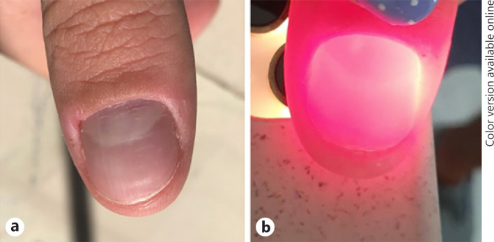
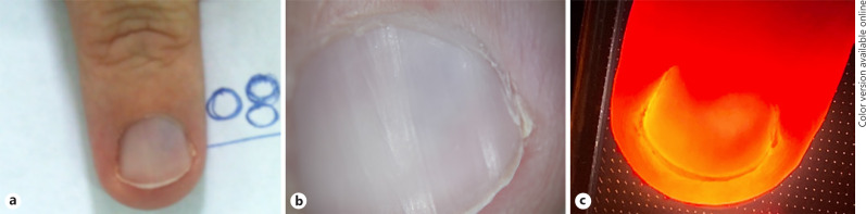
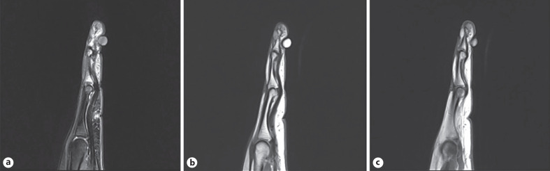
Diagnosis of subungual glomus tumors is mainly based on clinical symptoms, including paroxysmal pain, tenderness, and cold intolerance. Dermoscopy, ultrasonography, and MRI constitute further diagnostic tools, commonly performed to demarcate the tumor before surgery. Herein, we present 2 cases of subungual glomus tumors, which could be diagnosed after fingertip transillumination, highlighting that this technique can serve as an easy, noninvasive, and cost-effective adjuvant diagnostic tool, to facilitate the clinical diagnosis of subungual glomus tumors as well as their localization during preoperative assessment.
Keywords: Dermoscopy; Glomus tumor; Nail surgery; Nail tumors; Transillumination.
Copyright © 2021 by S. Karger AG, Basel.
Conflict of interest statement
The authors have no conflicts of interest to declare.
Figures
Similar articles
-
Eponychial flap elevation method for surgical excision of a subungual glomus tumor.Dermatol Ther. 2020 Nov;33(6):e14330. doi: 10.1111/dth.14330. Epub 2020 Oct 7. Dermatol Ther. 2020. PMID: 32975348
-
Outcome of transungual surgical approach with synthetic nail shield in the treatment of digital glomus tumors: A retrospective study.JPRAS Open. 2020 Oct 3;26:69-75. doi: 10.1016/j.jpra.2020.09.004. eCollection 2020 Dec. JPRAS Open. 2020. PMID: 33195786 Free PMC article.
-
Subungual glomus tumors of the hand: Treated by transungual excision.Indian J Orthop. 2015 Jul-Aug;49(4):403-7. doi: 10.4103/0019-5413.159611. Indian J Orthop. 2015. PMID: 26229160 Free PMC article.
-
Clinical and onychoscopic characteristics of subungual glomus tumor: a cross-sectional study.Int J Dermatol. 2021 Jun;60(6):693-702. doi: 10.1111/ijd.15358. Epub 2020 Dec 17. Int J Dermatol. 2021. PMID: 33332614 Review.
-
[Dermoscopy on subungual melanoma].Postepy Hig Med Dosw (Online). 2013 May 10;67:380-7. doi: 10.5604/17322693.1048815. Postepy Hig Med Dosw (Online). 2013. PMID: 23667097 Review. Polish.
Cited by
-
Blue Nail Discoloration: Literature Review and Diagnostic Algorithms.Am J Clin Dermatol. 2023 May;24(3):419-441. doi: 10.1007/s40257-023-00768-6. Epub 2023 Mar 27. Am J Clin Dermatol. 2023. PMID: 36971947 Review.
References
-
- Richert B, Lecerf P, Caucanas M, André J. Nail tumors. Clin Dermatol. 2013;31((5)):602–17. - PubMed
-
- Lipner SR, Scher RK. Longitudinal erythronychia of the fingernail. JAMA Dermatol. 2016;152((11)):1271–2. - PubMed
-
- Jakhar D, Kaur I. “Transillumination dermoscopy” for nail bed pathology. J Am Acad Dermatol. 2017 Dec 26 - PubMed
-
- Piraccini BM, Alessandrini A, Starace M. Onychoscopy: dermoscopy of the nails. Dermatol Clin. 2018;36((4)):431–8. - PubMed
Publication types
LinkOut - more resources
Full Text Sources
Other Literature Sources
