. 2021 Apr 20;6(17):11209-11222.
doi: 10.1021/acsomega.0c06232. eCollection 2021 May 4.

Distinct Modes of Tissue Expansion in Free Versus Earlier-Confined Boundaries for More Physiological Modeling of Wound Healing, Cancer Metastasis, and Tissue Formation

Affiliations

Distinct Modes of Tissue Expansion in Free Versus Earlier-Confined Boundaries for More Physiological Modeling of Wound Healing, Cancer Metastasis, and Tissue Formation

Abhimanyu Kiran et al. ACS Omega. .

Abstract

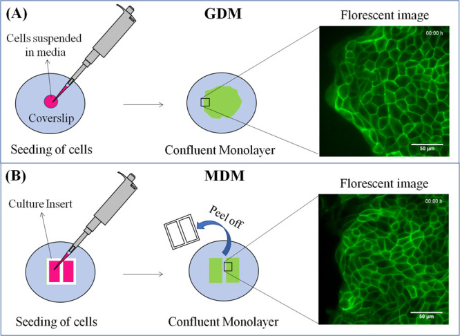
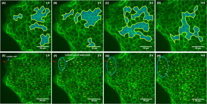
Collective cell migration is often seen in many biological processes like embryogenesis, cancer metastasis, and wound healing. Despite extensive experimental and theoretical research, the unified mechanism responsible for collective cell migration is not well known. Most of the studies have investigated artificial model wound to study the collective cell migration in an epithelial monolayer. These artificial model wounds possess a high cell number density compared to the physiological scenarios like wound healing (cell damage due to applied cut) and cancer metastasis (smaller cell clusters). Therefore, both systems may not completely relate to each other, and further investigation is needed to understand the collective cell migration in physiological scenarios. In an effort to fill this existing knowledge gap, we investigated the freely expanding monolayer that closely represented the physiological scenarios and compared it with the artificially created model wound. In the present work, we report the effect of initial boundary conditions (free and confined) on the collective cell migration of the epithelial cell monolayer. The expansion and migration aspects of the freely expanding and earlier-confined monolayer were investigated at the tissue and cellular levels. The freely expanding monolayer showed significantly higher expansion and lower migration in comparison to the earlier-confined monolayer. The expansion and migration rate of the monolayer exhibited a strong negative correlation. The study highlights the importance of initial boundary conditions in the collective cell migration of the expanding tissue and provides useful insights that might be helpful in the future to tune the collective cell migration in wound healing, cancer metastasis, and tissue formation.


Conflict of interest statement

The authors declare no competing financial interest.

Figures

Figure 1
Figure 1
Schematic representation of the monolayer formation. (A) GDM, (B) MDM.
Figure 2
Figure 2
Comparing the expansion of GDM and MDM. (A) Average expansion rate versus time, (B) average number of cells, (C) average cell area versus time, (D) representation of the number of cells with respect to change in their effective cell radius. Note: The black asterisk in (A–C) represents statistical significance obtained by applying an unpaired t-test between corresponding rows of GDM and MDM. The red asterisk in (A) represents statistical significance obtained by applying a paired t-test between the expansion rates of the same monolayer at a different time point. The black asterisk in (D) represents statistical significance obtained by applying an unpaired t-test between the similar effective cell radius of GDM and MDM. Standard errors plotted here are obtained from 5 independent experiments. The significant differences were determined by p-values, where *, **, and *** represents the significance level of p < 0.05, p < 0.01, and p < 0.001, respectively.
Figure 3
Figure 3
Comparison of migration between GDM and MDM. (A) Tracking of the cell center in GDM, (B) tracking of the cell center in MDM, (C) velocity field of GDM, (D) velocity field of MDM, (E) MSD as a function of time for GDM and MDM, (F) average migration rate versus time for GDM and MDM. Note: MSD vs time plot in (E) and standard errors in (F) are obtained from 5 independent experiments. The black asterisk represents statistical significance obtained by applying an unpaired t-test between corresponding rows of GDM and MDM. The significant differences were determined by p-values, where * and ** represent the significance level of p < 0.05 and p < 0.01, respectively.
Figure 4
Figure 4
Distribution of population and contribution of winner and loser cells in the overall expansion. (A) Percentage distribution of winner and loser cells in the overall population of the GDM, (B) percentage contribution of the winner and loser cells in the overall expansion of the GDM, (C) percentage distribution of winner and loser cells in the overall population of MDM, (D) percentage contribution of winner and loser cells in the overall expansion of MDM.
Figure 5
Figure 5
Comparison of expansion between winner and loser cells of GDM and MDM. (A) Net gain in the area by winner and loser cells, (B) average cell area versus time, (C) expansion rate of winner and loser cells of GDM, (D) expansion rate of winner and loser cells of MDM. Note: Standard error bars shown here are obtained from 5 independent experiments. The significance of paired and unpaired t-test was shown by the red and black asterisk, respectively. The significant differences were determined by p-values, where *, **, and *** represents the significance level of p < 0.05, p < 0.01, and p < 0.001, respectively.
Figure 6
Figure 6
Migration of winner and loser cells for GDM and MDM. (A) Velocity field of the winner (yellow vectors) and loser cells (red vectors) in GDM, (B) velocity field of the winner (yellow vectors) and loser cells (red vectors) in MDM, (C) MSD vs time graph for the winner and loser cells of GDM, (D) MSD vs time graph for winner and loser cells of MDM, (E) migration rate as a function of time for winner and loser cells of GDM and MDM, (F) temporal average migration rate of winner and loser cells of GDM and MDM.
Figure 7
Figure 7
Spatial comparison between GDM and MDM. (A) Average cell area vs cell rows, (B) average cell expansion rate vs cell rows, (C) average migration rate vs cell rows, (D) average expansion and migration rate of GDM as a function of cell rows, (E) average expansion and migration rate of MDM as a function of cell rows. Note: Standard error bars shown here were obtained from 5 independent experiments. The unpaired t-test was applied between the corresponding rows of GDM and MDM. The statistical significance of the unpaired t-test is shown by the black asterisk. The significant differences were determined by p-values, where *, **, and *** represents the significance level of p < 0.05, p < 0.01, and p < 0.001, respectively.
Figure 8
Figure 8
Morphological analysis of GDM and MDM. (A) Change in the cell shape with time, (B) SI as a function of the cell rows, (C) AR as a function of the cell rows. Note: The standard error bars shown here were obtained from 5 independent experiments. The unpaired t-test was applied between the corresponding rows of GDM and MDM. The statistical significance is shown by the black asterisk. The significant differences were determined by the p-values, where * and ** represent the significance level of p < 0.05 and p < 0.01, respectively.
Figure 9
Figure 9
3D interpretation of the 2D images. (A) Final image of the experiment is used as a reference image. The growing, shrinking, and fluctuating cells are represented by blue, red, and yellow circles, respectively. The ROIs “B”, “C” and “D” are marked in the reference image, (B) zoom-in of ROI “B,” and (C) zoom-in of ROI “C” contains the majority of growing cells. At t = 0 h, cells are focused on both planes. However, at t = 4 h, cells remained focused on the bottom plane and went out of focus on the top plane. Therefore, growing cells spread in the area by reducing in height and expanding in the area, (D) zoom-in of ROI “D”, the growing cells were out of focus in the top plane, whereas their neighboring cells (shrinking and fluctuating) remained in focus. It means that neighboring cells have more height than the growing cells. As these neighboring cells shrink or fluctuate with time, it may result from the push applied by the collectively growing or spreading cell colonies.
Figure 10
Figure 10
Dynamically growing cell colonies and the velocity field of GDM. The growing cell colonies found in (A) 1st h, (B) 2nd h, (C) 3rd h, and (D) 4th h. The velocity field of (E) 1st h, (F) 2nd h, (G) 3rd h, and (H) 4th h. Note: The growing cell colonies are represented by blue color and yellow outline. The blue ellipse shows the region of high collective cell alignment, and the red arrow represents the direction of motion of the leader cell.
Figure 11
Figure 11
Dynamically growing cell colonies and velocity field in MDM. The growing cell colonies in (A) 1st h, (B) 2nd h, (C) 3rd h, and (D) 4th hr. The velocity field in (E) 1st h, (F) 2nd h, (G) 3rd h, and (H) 4th hr. Note: The growing cell colonies are represented by blue color and yellow outline. The orange ellipse in (E) encloses the region having small magnitude vectors. The blue ellipse in (F–H) encloses the region showing high collective cell migration.

Similar articles

Cited by

References

    1. Friedl P.; Hegerfeldt Y.; Tusch M. Collective Cell Migration in Morphogenesis and Cancer. Int. J. Dev. Biol. 2004, 48, 441–449. 10.1387/ijdb.041821pf. - DOI - PubMed
    1. Martin P.; Parkhurst S. M. Parallels between Tissue Repair and Embryo Morphogenesis. Development 2004, 131, 3021–3034. 10.1242/dev.01253. - DOI - PubMed
    1. Lecaudey V.; Gilmour D. Organizing Moving Groups during Morphogenesis. Curr. Opin. Cell Biol. 2006, 18, 102–107. 10.1016/j.ceb.2005.12.001. - DOI - PubMed
    1. Friedl P. Prespecification and Plasticity: Shifting Mechanisms of Cell Migration. Curr. Opin. Cell Biol. 2004, 16, 14–23. 10.1016/j.ceb.2003.11.001. - DOI - PubMed
    1. De Pascalis C.; Etienne-Manneville S. Single and Collective Cell Migration: The Mechanics of Adhesions. Mol. Biol. Cell 2017, 28, 1833–1846. 10.1091/mbc.e17-03-0134. - DOI - PMC - PubMed

LinkOut - more resources