. 2021 Apr 28;6(18):12213-12222.
doi: 10.1021/acsomega.1c01058. eCollection 2021 May 11.

Improved Skin Permeability after Topical Treatment with Serine Protease: Probing the Penetration of Rapamycin by Scanning Transmission X-ray Microscopy

Affiliations

Improved Skin Permeability after Topical Treatment with Serine Protease: Probing the Penetration of Rapamycin by Scanning Transmission X-ray Microscopy

Gregor Germer et al. ACS Omega. .

Abstract

Drug penetration in human skin ex vivo following a modification of skin barrier permeability is systematically investigated by scanning transmission X-ray microscopy. Element-selective excitation is used in the O 1s regime for probing quantitatively the penetration of topically applied rapamycin in different formulations with a spatial resolution reaching <75 nm. The data were analyzed by a comparison of two methods: (i) two-photon energies employing the Beer-Lambert law and (ii) a singular value decomposition approach making use of the full spectral information in each pixel of the X-ray micrographs. The latter approach yields local drug concentrations more reliably and sensitively probed than the former. The present results from both approaches indicate that rapamycin is not observed within the stratum corneum of nontreated skin ex vivo, providing evidence for the observation that this high-molecular-weight drug inefficiently penetrates intact skin. However, rapamycin is observed to penetrate more efficiently the stratum corneum when modifications of the skin barrier are induced by the topical pretreatment with the serine protease trypsin for variable time periods ranging from 2 to 16 h. After the longest exposure time to serine protease, the drug is even found in the viable epidermis. High-resolution micrographs indicate that the lipophilic drug preferably associates with corneocytes, while signals found in the intercellular lipid compartment were less pronounced. This result is discussed in comparison to previous work obtained from low-molecular-weight lipophilic drugs as well as polymer nanocarriers, which were found to penetrate the intact stratum corneum exclusively via the lipid layers between the corneocytes. Also, the role of the tight junction barrier in the stratum granulosum is briefly discussed with respect to modifications of the skin barrier induced by enhanced serine protease activity, a phenomenon of clinical relevance in a range of inflammatory skin disorders.


Conflict of interest statement

The authors declare no competing financial interest.

Figures

Figure 1
Figure 1
X-ray absorption cross section of rapamycin in the O 1s regime. The inset (a) shows the O 1s → π* transition in greater detail. The structure of rapamycin is shown in the top right corner.
Figure 2
Figure 2
Penetration of rapamycin dissolved in ethanol (100 μg/cm2) topically applied to human SC ex vivo. Top: X-ray micrograph at 532.03 eV (white color corresponds to high transmission, black color to low transmission, see also included scheme of grayscale between the minimum (min) and maximum (max) of X-ray transmission), middle: rapamycin distribution as a function of skin depth at the same depth scale as the X-ray micrographs using 531.14 and 530.74 eV (approach 1, labeled ①); bottom: results from singular value decomposition (approach 2, labeled ②): (a) 10 min penetration time; (b) 100 min penetration time; and (c) 1000 min penetration time. The dashed thin white lines in the micrographs mark the top of the SC, that is, the skin surface is on the right-hand side. In (c), the top of the VE is also marked by another white dashed line. The scale bars correspond to 4 μm. The skin surface at the top edge of each micrograph is chosen as the reference of the depth scale. Horizontal black dashed lines are used to guide the eye for the depth profiles of rapamycin. The minimum (min) and maximum (max) correspond to (a) 80,000 and 175,000; (b) 68,500 and 179,500; (c) 67,000 and 127,500 counts/s, respectively.
Figure 3
Figure 3
Penetration of rapamycin dissolved in HEC gel (rapamycin: 425 μg/cm2 topically applied to the skin for 24 h). Top: X-ray micrographs at 532.03 eV; middle (approach 1, labeled ①): rapamycin distributions as a function of skin depth on the same length scale as the X-ray micrographs using the same photon energies as specified in Figure 2; bottom (approach 2, labeled ②): rapamycin distributions derived from singular value decomposition as a function of skin depth on the same length scale as the X-ray micrographs: (a) 2 h primary preparation with serine protease; (b) 4 h primary preparation with serine protease; (c) 8 h primary preparation with serine protease; and (d) 16 h primary preparation with serine protease. The vertical dashed thin white line on the right-hand side of each micrograph marks the skin surface. The left vertical dashed line corresponds to the top of the VE, which is located below the SC. The scale bar corresponds to 10 μm. The skin surface at the top edge of each micrograph is chosen as the reference of the depth scale. The dashed black horizontal lines in the rapamycin distributions as a function of depth are inserted to guide the eye. The minimum (min) and maximum (max) correspond to (a) 970,000 and 1,385,000; (b) 900,000 and 1,315,000; (c) 980,000 and 1,365,000; and (d) 942,500 and 1,445,000 counts/s, respectively.
Figure 4
Figure 4
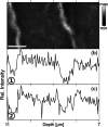
(a) High-resolution X-ray micrograph recorded at 532.03 eV in the SC of a skin sample exposed for 16 h to trypsin and subsequently for 1000 min to an ethanolic rapamycin solution. Lipid layers are marked by L and corneocytes by C. Pixel size: 30 nm2; the scale bar corresponds to 800 nm. The skin surface is chosen as the reference of the depth scale. (b) Integrated intensity of the rapamycin concentration on the same length scale as the micrograph using approach 1 (labeled ①) and (c) integrated intensity of the rapamycin concentration on the same length scale as the micrograph using approach 2 (labeled ②). The minimum (min) and maximum (max) correspond to 246,000 and 472,000 counts/s, respectively.

References

    1. Trommer H.; Neubert R. H. H. Overcoming the stratum corneum: The modulation of skin penetration—A review. Skin Pharmacol. Physiol. 2006, 19, 106–121. 10.1159/000091978. - DOI - PubMed
    1. Proksch E.; Brandner J. M.; Jensen J.-M. The skin: an indispensable barrier. Exp. Dermatol. 2008, 17, 1063–1072. 10.1111/j.1600-0625.2008.00786.x. - DOI - PubMed
    1. Elias P. M. Stratum corneum defensive functions: An integrated view. J. Invest. Dermatol. 2005, 125, 183–200. 10.1111/j.0022-202x.2005.23668.x. - DOI - PubMed
    1. Parisi R.; Symmons D. P. M.; Griffiths C. E. M.; Ashcroft D. M.; Identification M. Global Epidemiology of Psoriasis: A Systematic Review of Incidence and Prevalence. J. Invest. Dermatol. 2013, 133, 377–385. 10.1038/jid.2012.339. - DOI - PubMed
    1. Leung D. Y. M.; Boguniewicz M.; Howell M. D.; Nomura I.; Hamid Q. A. New insights into atopic dermatitis. J. Clin. Invest. 2004, 113, 651–657. 10.1172/jci21060. - DOI - PMC - PubMed

LinkOut - more resources