Novel Biomarkers for Diagnosing Periprosthetic Joint Infection from Synovial Fluid and Serum
- PMID: 34056503
- PMCID: PMC8154383
- DOI: 10.2106/JBJS.OA.20.00067
Novel Biomarkers for Diagnosing Periprosthetic Joint Infection from Synovial Fluid and Serum
Abstract
Background: Synovial fluid bacterial culture is the cornerstone of confirmation or exclusion of periprosthetic joint infection (PJI). The aim of this study was to assess synovial fluid and serum biomarker patterns of patients with total joint arthroplasty (TJA), and the association of these patterns with PJI.
Methods: Synovial fluid and serum samples were collected from 35 patients who were admitted to the Arthroplasty Unit of the Department of Orthopaedics and Traumatology at Turku University Hospital. Of the 25 patients who were included in the study, 10 healthy patients with an elective TJA for osteoarthritis served as the control group, and 15 patients who were admitted due to clinical suspicion of PJI with local redness, swelling, wound drainage, pain, and/or fever and who had a positive synovial fluid bacterial culture served as the study group. Logistic regression was used to assess the ability of 37 biomarkers (including cytokines, chemokines, and growth factors) with commercially available tests to detect PJIs.
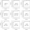
Results: In synovial fluid, the concentrations of sTNF-R1 and sTNF-R2 (soluble tumor necrosis factor receptors 1 and 2) and BAFF (B-cell activating factor, also known as TNFSF13B) were significantly higher in the PJI group (p < 0.002). In serum, the sTNF-R1 concentration was significantly higher in the PJI group, whereas the TWEAK (tumor necrosis factor-like weak inducer of apoptosis) and osteocalcin concentrations were significantly lower (p < 0.002). The sensitivity for detecting PJI using synovial fluid was 1.00 for sTNF-R2, 0.93 for sTNF-R1, and 0.87 for BAFF/TNFSF13B. The specificity of all 3 synovial markers was 1.00. The sensitivity using serum was 0.80 for TWEAK, 0.73 for sTNF-R1, and 0.80 for osteocalcin. The specificity of all 3 serum markers was 1.00.
Conclusions: Synovial sTNF-R2 is a promising new biomarker for detecting PJI. We are not aware of any previous reports of the use of sTNF-R2 in PJI diagnosis. More research is needed to assess the clinical importance of our findings.
Level of evidence: Diagnostic Level II. See Instructions for Authors for a complete description of levels of evidence.
Copyright © 2021 The Authors. Published by The Journal of Bone and Joint Surgery, Incorporated. All rights reserved.
Conflict of interest statement
Disclosure: The authors indicated that no external funding was received for any aspect of this work. The Disclosure of Potential Conflicts of Interest forms are provided with the online version of the article (http://links.lww.com/JBJSOA/A267).
Figures


Similar articles
-
Diagnosing periprosthetic joint infection: has the era of the biomarker arrived?Clin Orthop Relat Res. 2014 Nov;472(11):3254-62. doi: 10.1007/s11999-014-3543-8. Clin Orthop Relat Res. 2014. PMID: 24590839 Free PMC article.
-
Is synovial C-reactive protein a useful marker for periprosthetic joint infection?Clin Orthop Relat Res. 2014 Dec;472(12):3997-4003. doi: 10.1007/s11999-014-3828-y. Epub 2014 Jul 29. Clin Orthop Relat Res. 2014. PMID: 25070920 Free PMC article.
-
What is the performance of novel synovial biomarkers for detecting periprosthetic joint infection in the presence of inflammatory joint disease?Bone Joint J. 2021 Jan;103-B(1):32-38. doi: 10.1302/0301-620X.103B1.BJJ-2019-1479.R3. Bone Joint J. 2021. PMID: 33380185
-
Alpha-defensin-novel synovial fluid biomarker for the diagnosis of periprosthetic joint infection.Int Orthop. 2016 Dec;40(12):2447-2452. doi: 10.1007/s00264-016-3306-0. Epub 2016 Oct 7. Int Orthop. 2016. PMID: 27714447 Review.
-
Diagnostic Accuracy of Serum, Synovial, and Tissue Testing for Chronic Periprosthetic Joint Infection After Hip and Knee Replacements: A Systematic Review.J Bone Joint Surg Am. 2019 Apr 3;101(7):635-649. doi: 10.2106/JBJS.18.00632. J Bone Joint Surg Am. 2019. PMID: 30946198
Cited by
-
Comparison of the Effect of Intra-Articular, Periarticular, and Combined Injection of Analgesic on Pain Following Total Knee Arthroplasty: A Double-Blinded Randomized Clinical Trial.JB JS Open Access. 2022 Oct 7;7(4):e22.00074. doi: 10.2106/JBJS.OA.22.00074. eCollection 2022 Oct-Dec. JB JS Open Access. 2022. PMID: 36226033 Free PMC article.
-
Synovial Fluid Interleukin Levels Cannot Distinguish between Prosthetic Joint Infection and Active Rheumatoid Arthritis after Hip or Knee Arthroplasty.Diagnostics (Basel). 2022 May 11;12(5):1196. doi: 10.3390/diagnostics12051196. Diagnostics (Basel). 2022. PMID: 35626351 Free PMC article.
-
Serum and Synovial Markers in Patients with Rheumatoid Arthritis and Periprosthetic Joint Infection.J Pers Med. 2022 May 17;12(5):810. doi: 10.3390/jpm12050810. J Pers Med. 2022. PMID: 35629231 Free PMC article.
-
Enhanced Diagnostic Accuracy for Septic Arthritis Through Multivariate Analysis of Serum and Synovial Biomarkers.J Clin Med. 2025 Aug 1;14(15):5415. doi: 10.3390/jcm14155415. J Clin Med. 2025. PMID: 40807041 Free PMC article.
-
Synovial calprotectin for the diagnosis of periprosthetic joint infection: a diagnostic meta-analysis.J Orthop Surg Res. 2022 Jan 4;17(1):2. doi: 10.1186/s13018-021-02746-2. J Orthop Surg Res. 2022. PMID: 34983582 Free PMC article.
References
-
- Parvizi J, Gehrke T; International Consensus Group on Periprosthetic Joint Infection. Definition of periprosthetic joint infection. J Arthroplasty. 2014. July;29(7):1331. Epub 2014 Mar 21. - PubMed
-
- Della Valle CJ, Sporer SM, Jacobs JJ, Berger RA, Rosenberg AG, Paprosky WG. Preoperative testing for sepsis before revision total knee arthroplasty. J Arthroplasty. 2007. September;22(6)(Suppl 2):90-3. Epub 2007 Jul 26. - PubMed
-
- Squire MW, Della Valle CJ, Parvizi J. Preoperative diagnosis of periprosthetic joint infection: role of aspiration. AJR Am J Roentgenol. 2011. April;196(4):875-9. - PubMed
LinkOut - more resources
Full Text Sources
Other Literature Sources
Research Materials
