. 2021 Apr 7;3(2):fcab063.
doi: 10.1093/braincomms/fcab063. eCollection 2021.

Dominant ACO2 mutations are a frequent cause of isolated optic atrophy

Affiliations

Dominant ACO2 mutations are a frequent cause of isolated optic atrophy

Majida Charif et al. Brain Commun. .

Abstract

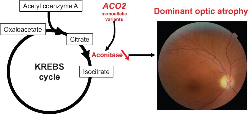
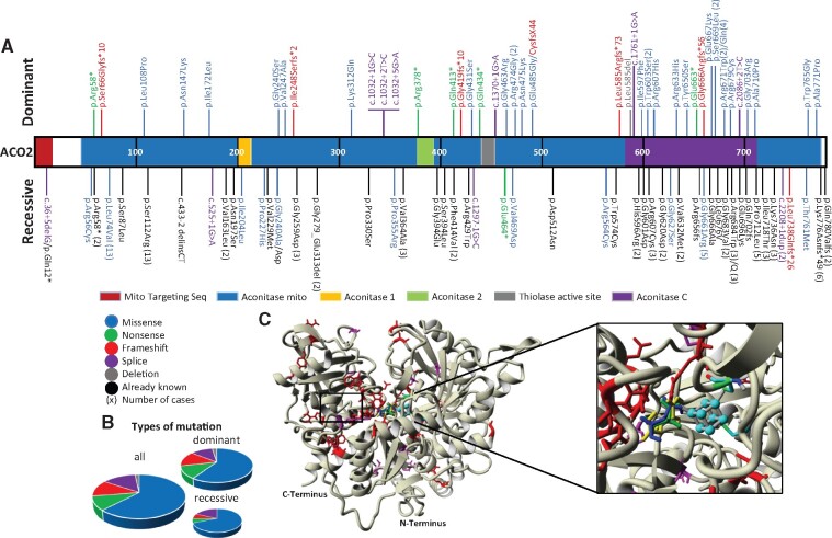
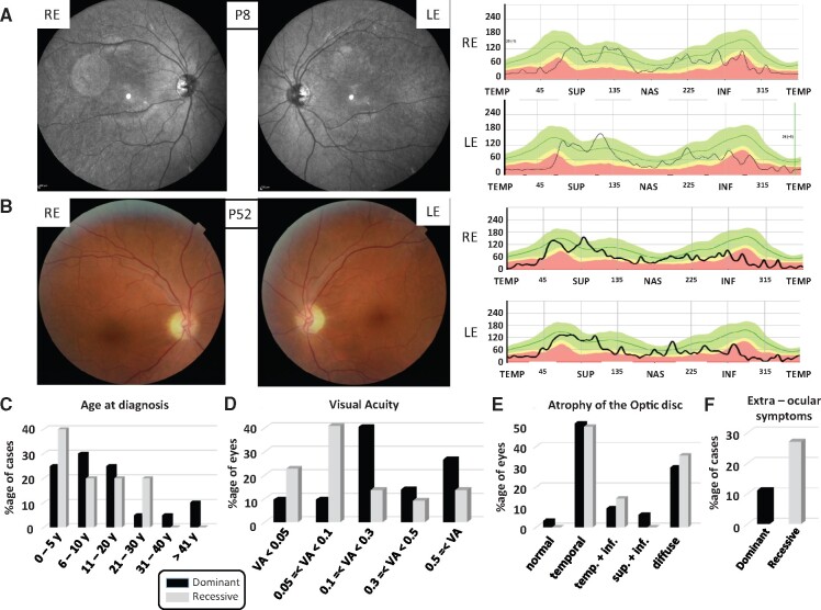
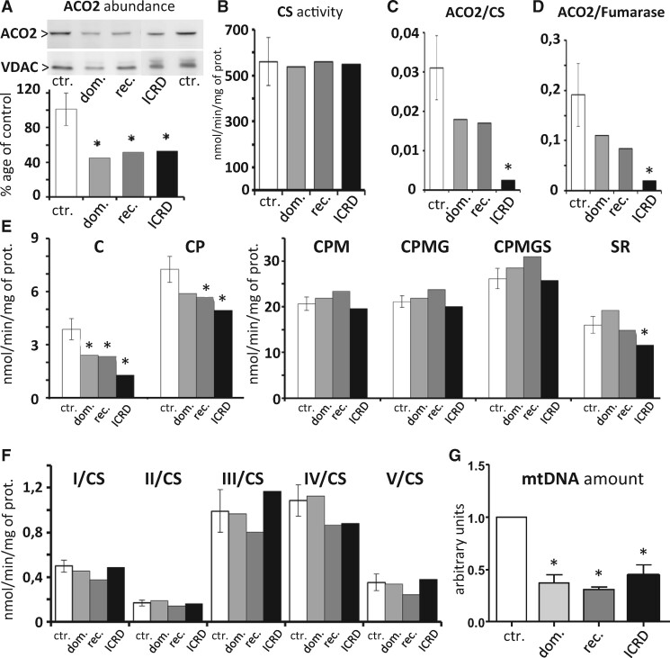
Biallelic mutations in ACO2, encoding the mitochondrial aconitase 2, have been identified in individuals with neurodegenerative syndromes, including infantile cerebellar retinal degeneration and recessive optic neuropathies (locus OPA9). By screening European cohorts of individuals with genetically unsolved inherited optic neuropathies, we identified 61 cases harbouring variants in ACO2, among whom 50 carried dominant mutations, emphasizing for the first time the important contribution of ACO2 monoallelic pathogenic variants to dominant optic atrophy. Analysis of the ophthalmological and clinical data revealed that recessive cases are affected more severely than dominant cases, while not significantly earlier. In addition, 27% of the recessive cases and 11% of the dominant cases manifested with extraocular features in addition to optic atrophy. In silico analyses of ACO2 variants predicted their deleterious impacts on ACO2 biophysical properties. Skin derived fibroblasts from patients harbouring dominant and recessive ACO2 mutations revealed a reduction of ACO2 abundance and enzymatic activity, and the impairment of the mitochondrial respiration using citrate and pyruvate as substrates, while the addition of other Krebs cycle intermediates restored a normal respiration, suggesting a possible short-cut adaptation of the tricarboxylic citric acid cycle. Analysis of the mitochondrial genome abundance disclosed a significant reduction of the mitochondrial DNA amount in all ACO2 fibroblasts. Overall, our data position ACO2 as the third most frequently mutated gene in autosomal inherited optic neuropathies, after OPA1 and WFS1, and emphasize the crucial involvement of the first steps of the Krebs cycle in the maintenance and survival of retinal ganglion cells.

Keywords: ACO2; Krebs cycle; aconitase 2; mitochondria; optic neuropathy.


Figures

Graphical Abstract
Graphical Abstract
Figure 1
Figure 1
Localization and quantification of the ACO2 variants on the protein primary structure. (A) The structure of the ACO2 protein including the different functional domains is represented, with all the dominant mutations identified shown on top, and all the recessive mutations identified shown below (different colours according to the type of variant), interspersed with the published recessive variants (in black). The number of times that a variant has been identified is indicated between brackets. (B) Representation of the different variant subtypes affecting ACO2 in all optic atrophy cases (left), in dominant cases (right top) and in recessive cases (right bottom). There are no significant differences (P-value: 0.80) between dominant and recessive groups. (C) Three-dimensional structure of ACO2 protein showing the dominant (red) and recessive (magenta) variants and the ligands localization. [4Fe, 4S] cluster (cyan), citrate (green), cis-aconitate (blue) and isocitrate (yellow).
Figure 2
Figure 2
Clinical data of the dominant and recessive ACO2 individuals. (A and B) Left: Eye fundus pictures of individuals with a dominant (A: P8) and a recessive (B: P52) ACO2 mutation revealing the temporal pallor of the optic discs in both REs and LEs. Right: Evaluation of the retinal nerve fibre layer by optic coherence tomography in the same individuals, disclosing in both cases the retinal nerve fibre layer reduction in the temporal quadrants. The green area defines the 5th to 95th, the yellow area the 1st to 5th and the red area below the 1st percentiles. INF = inferior quadrants; LE = left eye; NAS = nasal; RE = right eye; SUP = superior; TEMP = temporal. (C) Ages at diagnosis categorized in six groups and represented as percentages of the number of individuals in the dominant and recessive cohorts (y = years) are not significantly different (P-value: 0.54). (D) Visual acuities (VA), categorized in five ranges, and represented as percentages of the total number of eyes are significantly different between dominant and recessive cases (P-value: 0.001). (E) Alterations of the optic disc, according to the different retinal nerve fibre layer quadrants were assessed by optical coherence tomography. Data are represented as percentages of each alteration from all eyes for which the data were available. (temp. = temporal; inf. = inferior; sup. = superior) and are not significantly different between dominant and recessive cases (P-value: 0.88). (F) Percentage of dominant and recessive ACO2 individuals presenting extra-ocular symptoms.
Figure 3
Figure 3
Analysis of dominant and recessive ACO2 mutated fibroblasts, compared to an ACO2-related ICRD fibroblast cell line. All experiments were performed at least in two independent replicates for each control (ctr.) and dominant (dom.) and recessive (rec.) ACO2 fibroblasts, and compared to one ACO2-related ICRD fibroblast cell line (ICRD). Results are mean ± SD. Statistical analysis of results from all the following experiments was performed using the two-tailed paired t-test. (A) Western blots with antibodies against ACO2 and VDAC proteins. The quantification of the relative ratio shows a significant decrease of ACO2 protein in all patient fibroblasts (*P-value <0.05). (B) CS activity is not affected by ACO2 mutations. (C and D) Relative ACO2 activity normalized to the CS (C) and to the fumarase (D) activities shows a tendency to decrease in the dominant and recessive ACO2 fibroblasts, and a significant decrease in the ICRD fibroblasts (*P-value <0.05). (E) The assessment of fibroblast respiration (mitochondrial oxygen rates related to maximal phosphorylation condition in permeablized fibroblasts) by oxygraphy, using the Krebs cycle substrates, Citrate (C), pyruvate (P), malate (M), glutamate (G), succinate (S), followed by the inhibition of complex I by rotenone (R), show that the respiration related to the use of citrate is decreased in all ACO2 fibroblasts, partially increased by pyruvate, and fully restored by malate. Further stimulation by glutamate and succinate is limited, and only the ICRD fibroblasts are significantly more affected than the other fibroblasts by the Rotenone (*P-value <0.05). (F) Enzymatic activities from four independent experiments of the respiratory complexes (CI to CV) from the control and the ACO2 mutated fibroblast strains related to the CS enzymatic activity did not reveal a significant difference between control and mutated fibroblasts. (G) Mitochondrial DNA copy number in ACO2 mutated fibroblasts normalized to control fibroblasts reveals a significant decrease in the mitochondrial genome in all ACO2 cells (*P-value <0.05).

References

    1. La Morgia C, Maresca A, Caporali L, Valentino ML, Carelli V.. Mitochondrial diseases in adults. J Intern Med. 2020;287(6):592–608. - PubMed
    1. Martius C.Über den Abbau der Citronensäure. Zschr Physiol Chem. 1937;247:104–110.
    1. Fukada M, Yamada K, Eda S, et al.Identification of novel compound heterozygous mutations in ACO2 in a patient with progressive cerebral and cerebellar atrophy. Mol Genet Genomic Med. 2019;7(7):e00698. - PMC - PubMed
    1. Sharkia R, Wierenga KJ, Kessel A, et al.Clinical, radiological, and genetic characteristics of 16 patients with ACO2 gene defects: Delineation of an emerging neurometabolic syndrome. J Inherit Metab Dis. 2019;42(2):264–275. doi: 10.1002/jimd.12022. Epub 28 Jan 2019. PMID: 30689204. - PubMed
    1. Spiegel R, Pines O, Ta-Shma A, et al.[Database] Infantile cerebellar-retinal degeneration associated with a mutation in mitochondrial aconitase ACO2. Am J Hum Genet. 2012;90(3):518–523. - PMC - PubMed

LinkOut - more resources