. 2021 May 24;1(5):669-689.
doi: 10.1021/jacsau.1c00025. Epub 2021 Apr 15.

A Class of Valuable (Pro-)Activity-Based Protein Profiling Probes: Application to the Redox-Active Antiplasmodial Agent, Plasmodione

Affiliations

A Class of Valuable (Pro-)Activity-Based Protein Profiling Probes: Application to the Redox-Active Antiplasmodial Agent, Plasmodione

Bogdan Adam Cichocki et al. JACS Au. .

Abstract

Plasmodione (PD) is a potent antimalarial redox-active drug acting at low nM range concentrations on different malaria parasite stages. In this study, in order to determine the precise PD protein interactome in parasites, we developed a class of (pro-)activity-based protein profiling probes (ABPP) as precursors of photoreactive benzophenone-like probes based on the skeleton of PD metabolites (PDO) generated in a cascade of redox reactions. Under UV-photoirradiation, we clearly demonstrate that benzylic oxidation of 3-benzylmenadione 11 produces the 3-benzoylmenadione probe 7, allowing investigation of the proof-of-concept of the ABPP strategy with 3-benzoylmenadiones 7-10. The synthesized 3-benzoylmenadiones, probe 7 with an alkyne group or probe 9 with -NO2 in para position of the benzoyl chain, were found to be the most efficient photoreactive and clickable probes. In the presence of various H-donor partners, the UV-irradiation of the photoreactive ABPP probes generates different adducts, the expected "benzophenone-like" adducts (pathway 1) in addition to "benzoxanthone" adducts (via two other pathways, 2 and 3). Using both human and Plasmodium falciparum glutathione reductases, three protein ligand binding sites were identified following photolabeling with probes 7 or 9. The photoreduction of 3-benzoylmenadiones (PDO and probe 9) promoting the formation of both the corresponding benzoxanthone and the derived enone could be replaced by the glutathione reductase-catalyzed reduction step. In particular, the electrophilic character of the benzoxanthone was evidenced by its ability to alkylate heme, as a relevant event supporting the antimalarial mode of action of PD. This work provides a proof-of-principle that (pro-)ABPP probes can generate benzophenone-like metabolites enabling optimized activity-based protein profiling conditions that will be instrumental to analyze the interactome of early lead antiplasmodial 3-benzylmenadiones displaying an original and innovative mode of action.


Conflict of interest statement

The authors declare no competing financial interest.

Figures
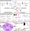
Figure 1
Figure 1
(A) Bioactivation of plasmodione (PD 1): Upon internalization in the parasite, plasmodione PD (1) is proposed to generate the drug metabolite PDOox (2) by benzylic oxidation (step ①), the 3-benzoylmenadione (benzoylMD), which, under its oxidized form, possesses a photoreactive benzophenone-like moiety (indicated in red). This metabolite is further reduced (step ②) and under its reduced form (3) takes part in oxidoreductase-mediated redox-cycling (step ③), leading to ROS-induced parasite death. In addition, the generation of a third metabolite, namely the benzo[c]xanthen-7-one (benzoxanthone, PDO-BX) derivative (4), has been envisioned as one possible metabolite generated through an oxidative phenolic coupling reaction from the 1-electron-reduced benzoylMD (3) radical (step ④). Significantly, PDOred (3) exists in different mesomeric species, but for the clarity of the scheme, only one species is shown. (B) Plasmodione-activity based probes (PD-ABPP): The common scaffold of the PD-ABPP probes 711 is a photoreactive 3-benzoylmenadione, functionalized by different electron-withdrawing groups in para-position (-CF3 or -NO2 or -alkyne) affecting their photoreactivity. 3-Benzylmenadiones (such as 1, 5, 11) are not photoreactive per se, while probes 611 in the benzoylMD series are photoreactive. (C) ABPP strategy: This approach is aimed at identifying drug activity-based protein profiling in living parasites incubated with a parent PD-ABPP precursor designed as (pro-)PD-ABPP. The (pro-)PD-ABPP probe is released upon bioactivation through the benzylic oxidation. In this paper, our aim is to showcase the proof-of-concept by starting the work from the benzoylmenadione derivatives (metabolites and ABPP probes). Upon UV irradiation, covalent cross-linking of PD-ABP to its potential targets is expected to occur. Further, a reporter click reaction between the probe-derived alkyne and the fluorescent rhodamine azide or biotin azide reveals successful cross-linking of probe to peptides and GRs as protein models, which can be analyzed by SDS PAGE and/or LC-MS/MS. Identification of the site where the ABPP probe was bound to both GRs was investigated and discussed.
Figure 2
Figure 2
Mass spectrometric analysis of the photochemical reaction mixtures. Field-desorption mass spectrometry (FD-MS) analyses of the photochemical reaction mixtures of (panel A) the 3-benzoylmenadione 6 or (panel B) the 3-benzylmenadione 5 derivatives, in the presence of the diprotected methionine nMet.
Scheme 1
Scheme 1. Synthesis of the 3-Benzoylmenadiones 710 (Paths A and B) through the Friedel–Crafts Reaction Variant and the 3-Benzylmenadione 11 (Path C) through the Kochi–Anderson Reaction,
Reaction conditions: (i) 1. SnCI2 cc HCI, EtOH, rt, 2 h, 2. Me2SO4 acetone, KOH, MeOH, 60 °C, 4 h; (ii) TfOH/TFAA, DCM, and A. 4-iodobenzoic acid 7a, or 3-iodo-4-(trifluoromethyl)benzoic acid 8a, B. 4-nitro-3-fluorobenzoic acid 9a, or 3-fluoro-4-(trifluoromethyl)benzoic acid 10a, 0 °C then rt, 16 h; (iii) Cul, Pd(PPh3)2CI2 NEt3 rt, 16 h, HC ≡ C-TMS; (iv) TBAF, THF, rt, 1.5 h; (v) CAN, H2O/MeCN, rt, 3 h; (vi) K2CO3 DMF, propargylic alcohol, 60 °C, 24 h; (vii) 4-iodophenylacetic acid, AgNO3 Na2S2O8 MeCN/H2O, reflux, 4 h.
Figure 3
Figure 3
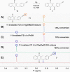
Overlay of the 1H NMR spectra in the 2.8–3.4 ppm area of (A) pure probe 11 and reaction mixtures of 3-benzylmenadione probe 11 in various solvents after 72 h of UV-photoirradiation at 350 nm: (B) in a 1:1 H2O:MeCN system; (C) in iPrOH; (D) in a 1:1 CH2Cl2:iPrOH system; (E) pure 3-benzoylmenadione probe 7.
Figure 4
Figure 4
Phosphate buffer affects the click reaction efficiency. An increase of CuSO4 and THPTA ratios and decrease of PBS concentrations led to a click reaction between probe 7 and RA as efficient as in pure water. Left panel: overnight click reaction of RA with probe 7 in 47 mM or 12 mM phosphate buffer. Copper-ligand preincubated mixture was added after 40 min of incubation. Copper-ligand preincubation mixture – 1:1 = 132 μM of TCEP, CuSO4, and THPTA; 5:5 = 132 μM of TCEP and 660 μM of CuSO4 and THPTA. Chromatograms using absorption detection at 507 nm are shown. The two peaks evidenced for RA are related to both isomers in solution. Right panel: Yields of reactions determined from reactions in left panel; additional reaction data in H2O and 24 mM PBS are shown. Reactions were analyzed by LC-MS. Total area of rhodamine absorption at 507 nm of the peaks corresponding to the product mass was measured and normalized to 24 μM RA unreacted control. N = 3 independent experiments Error bars represent ± SD.
Figure 5
Figure 5
Probe 9 forms photoadducts with GSH in aqueous ACN conditions. (A) Chromatogram using absorption detection at 200–600 nm obtained by LC-MS analysis of reaction mixture containing GSH (3 mM) without ABPP probe upon 8 min UV irradiation. Glutathione disulfide (GSSG, RT = 4 min) is formed in the reaction by oxidation of GSH (RT = 4.25). (B) Under the conditions described in (A), 200–600 nm chromatogram is depicted after LC-MS analysis of reaction mixture containing probe 9 (600 μM) and 3 mM GSH upon UV irradiation for 8 min (n = 4). Multiple peaks corresponding to different GSH and GSSG adducts (different cross-linking site, GSH and GSSG fragments, double cross-linking) are visible in the chromatograms. Peak corresponding to mass of photo-cross-linked adduct of full GSH and probe 9 is highlighted in red box (RT = 33.5 min). (C) Left panel – Extracted ion chromatogram of m/z = 618.16 Da from reaction in (B). Right panel – Fragmentation pattern of the selected peak in (B) spectrum showing adduct m/z at 681.16 Da. ECG – amino acid letter code of GSH. Peaks on the right side from m/z = 308.08 originate from probe 9-derived BX-SG fragmentation, on the left side from GSH fragmentation.
Scheme 2
Scheme 2. Chemical Analysis of the Insertion Products upon Photoirradiation of the ABPP Probe 9 with Glutathione (GSH) by Mass Spectrometry
Two pathways of photoreactivity of the benzoylmenadione were expressed through the formation of two insertion products (blue box) with nucleophilic partners.
Figure 6
Figure 6
Photoreduction of probe 9 generates multiple probe-insertion products with GSH. (A) 280 nm UV-chromatogram overlaid with extracted ion chromatograms (EIC) corresponding to detected adduct and probe species. Reaction analyzed after 10 min of UV-irradiation with GSH and probe 9 (RT = 39 min). Red box indicates position of 2-(S-methyl) adduct peak (RT = 23 min). (B) 280 nm UV-chromatogram and EICs for an overnight photoreaction of probe 9 with GSH. Red box indicates position of 9-BX adduct (RT = 26.5 min). (C) MS/MS spectra of probe 9 peak (RT = 39 min; left panel) and 9-BX peak (RT = 40.5 min; right panel) from the reaction depicted in B).
Scheme 3
Scheme 3. Mechanism of Formation of Both Observed Insertion Products (Blue Box) via Pathway 3 upon Photoirradiation of the ABPP Probe 9 with Glutathione
The structure of the intermediate 2-(SG-methyl)-probe 9 adduct, formed after 10 min-irradiation, was deduced by ESI-MS/MS mass spectrometry.
Figure 7
Figure 7
Probe 9 cross-links to hGR (A) MS/MS fragmentation pattern of identified peptides of hGR photoreaction mixture with probe 9. Left panel – peptide cross-linked at K397 (2893.23 Da = Y393–K416 + 9 - 18 Da; dehydration is common for benzophenone adducts). Right panel – peptide cross-linked at C234 (2874.23 + 9-BX[NH2] Da). Red circles indicate identified cross-linking sight. (B) Left panel – position of K256–7, K397 (blue), C234 (orange), Tyr197 (pink), and FAD (yellow) have been marked on the previously reported hGR dimer structure cross-linked to menadione analogue (red). The substrate binding cleft leading to the catalytic disulfide bridge is visible between K397 and menadione core (orange triangle). (C) Magnification on C234 (yellow) containing the binding pocket with indicated water molecules (violet balls). Surface of A241 (blue) and H374 (pink) at pocked opening and V200 (green) in cavity is visible.
Figure 8
Figure 8
Probe 9 cross-links to PfGR. (A) Positions of cross-linked amino acids (K415 – blue, V6 – pink) to probe 9 in the PfGR structure. The substrate binding cleft leading to the catalytic disulfide bridge and interspace cavity opening is indicated by the orange triangles. (B) Images picturing the distance between cross-linked K397 in hGR (left panels), K415 in PfGR (right panels), and the interspace cavity opening. Upper panel – amino acid positions on protein chain. Lower panels – surface density of cavity opening. Blue – cross-linked lysine; Green and violet – amino acids of the cavity opening.
Figure 9
Figure 9
Pull-down of hGR labeled with ABPP probes 7 and 9 and clicked with biotin tag. SDS-PAGE gel stained with Coomassie is pictured. For each reaction, 2% of the reaction before pulldown and 50% of the elution after avidin binding were loaded on the gel. hGR is localized at the height of the 55 kDa marker band. M – marker.
Figure 10
Figure 10
ESI-MS and CID-MS analysis of PDO-BX 4-heme complexes. (A) ESI mass spectrum (exit potential: 120 V) of a 1:1 mixture of 50 μM heme and 50 μM PDO-BX 4 in H2O/CH3CN (5/95) – 1% formic acid. (B) Stability responses of the BX 4-heme (at m/z = 960.2 and at 975.3), AQ-heme (at m/z = 971.3) and CQ-heme (at m/z = 935.4) complexes obtained by CID-MS experiments. ESI-MS+; 120 V < fragmentor< 400 V with 20 V increments. (C) Proposed molecular structure of ironIII-hematin species alkylated by the BX 4 and comparison between simulated and observed mass signatures of species A and D.

References

    1. Galmozzi A.; Dominguez E.; Cravatt B. F.; Saez E.. Application of Activity-Based Protein Profiling to Study Enzyme Function in Adipocytes. In Methods in Enzymology; Academic Press: San Diego, 2014; pp 151–169. - PMC - PubMed
    1. Whitby L. R.; Obach R. S.; Simon G. M.; Hayward M. M.; Cravatt B. F. Quantitative Chemical Proteomic Profiling of the in Vivo Targets of Reactive Drug Metabolites. ACS Chem. Biol. 2017, 12, 2040–2050. 10.1021/acschembio.7b00346. - DOI - PMC - PubMed
    1. Liu Y.; Patricelli M. P.; Cravatt B. F. Activity-Based Protein Profiling: The Serine Hydrolases. Proc. Natl. Acad. Sci. U. S. A. 1999, 96, 14694–14699. 10.1073/pnas.96.26.14694. - DOI - PMC - PubMed
    1. Yoo E.; Schulze C. J.; Stokes B. H.; Onguka O.; Yeo T.; Mok S.; Gnädig N. F.; Zhou Y.; Kurita K.; Foe I. T.; Terrell S. M.; Boucher M. J.; Cieplak P.; Kumpornsin K.; Lee M. C. S.; Linington R. G.; Long J. Z.; Uhlemann A. C.; Weerapana E.; Fidock D. A.; Bogyo M. The Antimalarial Natural Product Salinipostin A Identifies Essential α/β Serine Hydrolases Involved in Lipid Metabolism in P. Falciparum Parasites. Cell Chem. Biol. 2020, 27, 143–157. 10.1016/j.chembiol.2020.01.001. - DOI - PMC - PubMed
    1. Rostovtsev V. V.; Green L. G.; Fokin V. V.; Sharpless K. B. A Stepwise Huisgen Cycloaddition Process: Copper(I)-Catalyzed Regioselective “Ligation” of Azides and Terminal Alkynes. Angew. Chem., Int. Ed. 2002, 41, 2596–2599. 10.1002/1521-3773(20020715)41:14<2596::AID-ANIE2596>3.0.CO;2-4. - DOI - PubMed

LinkOut - more resources