. 2021 May 24;1(5):690-696.
doi: 10.1021/jacsau.1c00006. Epub 2021 Apr 23.

A General Method to Improve Fluorophores Using Deuterated Auxochromes

Affiliations

A General Method to Improve Fluorophores Using Deuterated Auxochromes

Jonathan B Grimm et al. JACS Au. .

Abstract

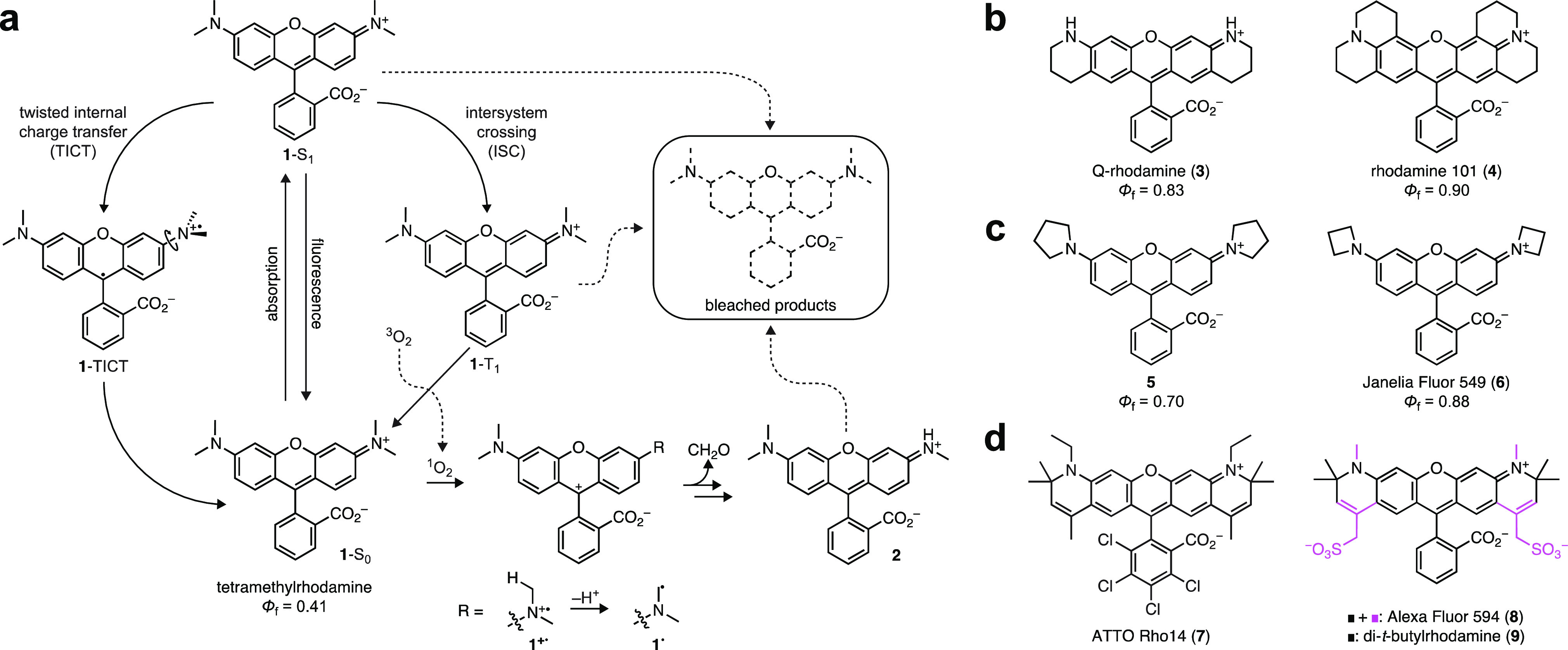
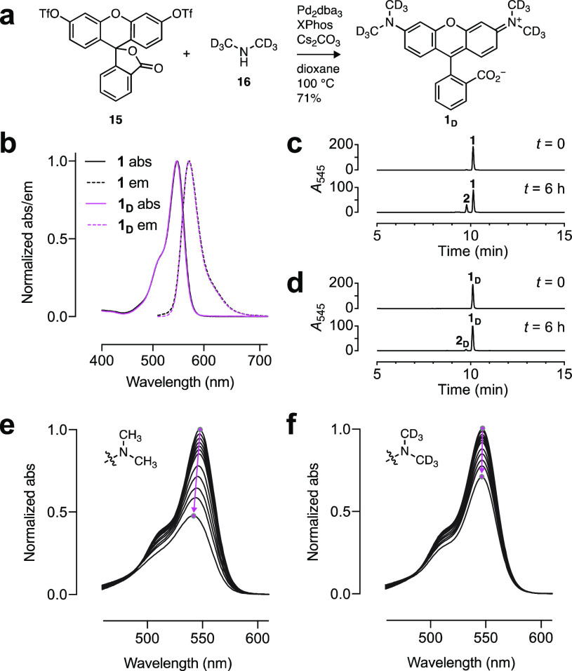
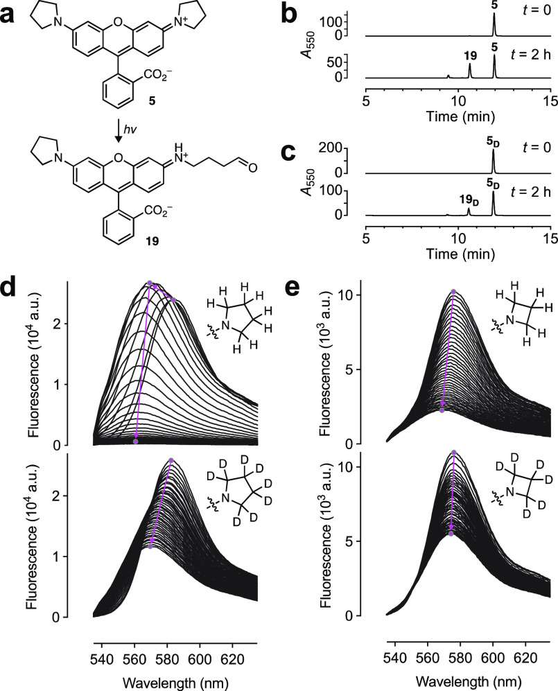
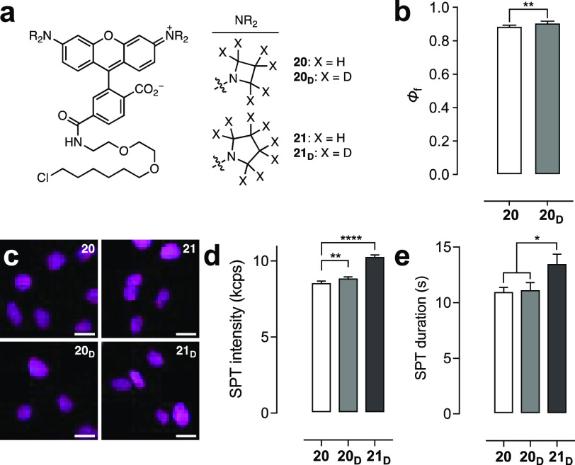
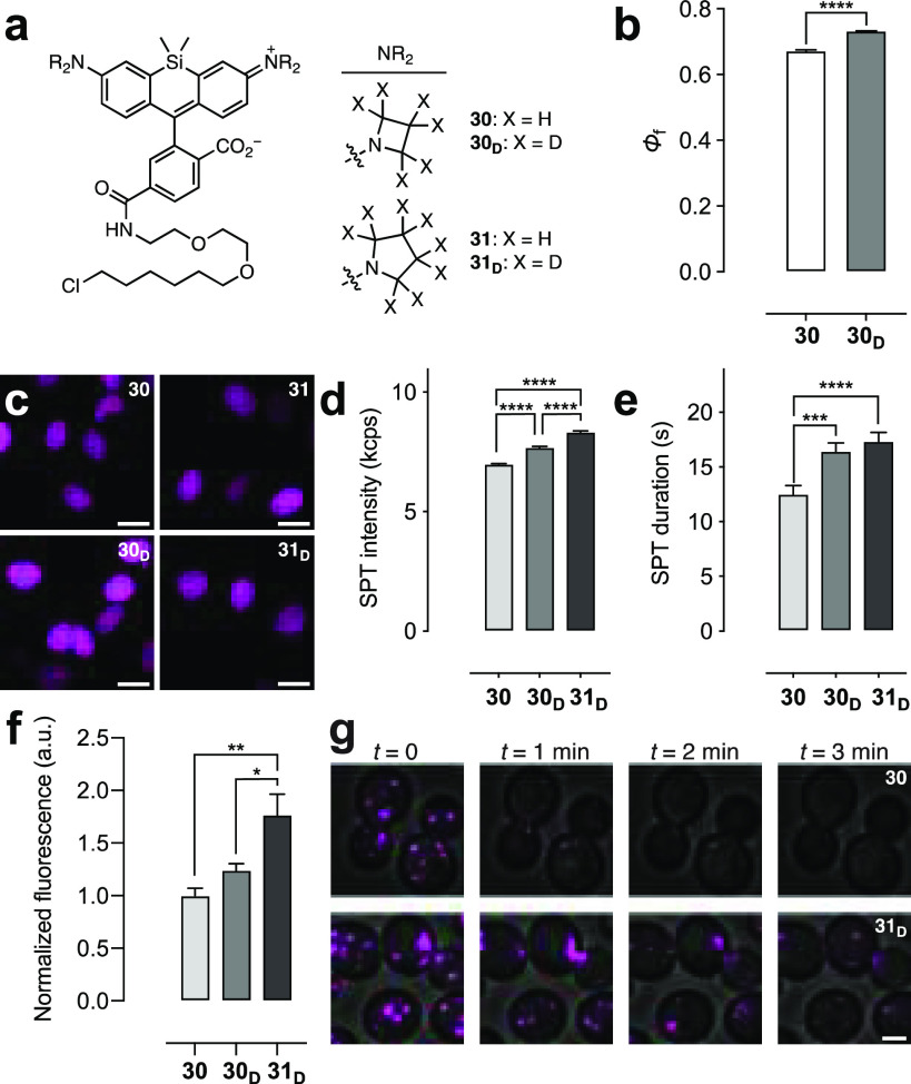
Fluorescence microscopy relies on dyes that absorb and then emit photons. In addition to fluorescence, fluorophores can undergo photochemical processes that decrease quantum yield or result in spectral shifts and irreversible photobleaching. Chemical strategies that suppress these undesirable pathways-thereby increasing the brightness and photostability of fluorophores-are crucial for advancing the frontier of bioimaging. Here, we describe a general method to improve small-molecule fluorophores by incorporating deuterium into the alkylamino auxochromes of rhodamines and other dyes. This strategy increases fluorescence quantum yield, inhibits photochemically induced spectral shifts, and slows irreparable photobleaching, yielding next-generation labels with improved performance in cellular imaging experiments.


Conflict of interest statement

The authors declare the following competing financial interest(s): Patents and patent applications describing azetidine- and deuterium-containing fluorophores (with inventors J.B.G. and L.D.L.) are assigned to HHMI.

Figures

Figure 1
Figure 1
Photophysics of rhodamines and methods to improve rhodamine properties. (a) Photophysics of tetramethylrhodamine (TMR, 1). (b) Structures of rigidified rhodamines 34. (c) Structures of cyclic amine-containing rhodamines 56. (c) Structures of α-quaternary rhodamines 79.
Figure 2
Figure 2
Deuterated tetramethylrhodamine. (a) Synthesis of 1D. (b) Normalized absorption (abs) and fluorescence emission (em) spectra of 1 and 1D. (c, d) LC–MS traces of 1 (c) and 1D (d) before and after photobleaching using 560 nm (1.02 W/cm2, 6 h). (e, f) Sequential absorption spectra of 1 (e) and 1D (f) during photobleaching using 560 nm (1.02 W/cm2). The magenta arrows highlight the shift in λabs and absorption intensity over time.
Figure 3
Figure 3
Photostability and chromostability of 5, 5D, 6, and 6D. (a) Photochemical dealkylation of 5 to form aldehyde 19. (b, c) LC–MS traces of 5 (b) and 5D (c) before and after photobleaching. (d, e) Sequential fluorescence emission spectra of (d) 5 and 5D or (e) 6 and 6D during photobleaching using a 532 nm laser (0.96 W/cm2; 40 spectra taken over 40 min). The magenta arrows highlight the shift in λem and intensity over time.
Figure 4
Figure 4
Performance of rhodamine ligands. (a) Structures of HaloTag ligands 20, 20D, 21, and 21D. (b) Φf of HaloTag protein conjugates of 20 and 20D. (c) Confocal microscopy images of live U2OS cells expressing HaloTag–histone H2B incubated with HaloTag ligands 20, 20D, 21, and 21D (200 nM, 2 h); ex/em = 561 nm/565–632 nm; scale bars: 21 μm. (d, e) SPT intensity (kilocounts per second, kcps) (d) or duration (s) (e) from cells labeled with 20, 20D, or 21D. All error bars: SEM.
Figure 5
Figure 5
Performance of Si–rhodamine ligands. (a) Structures of HaloTag ligands 30, 30D, 31, and 31D. (b) Φ of HaloTag protein conjugates of 30 or 30D. (c) Confocal microscopy images of live U2OS cells expressing HaloTag–histone H2B incubated with HaloTag ligands 30, 30D, 31, and 31D (200 nM, 2 h); ex/em = 640 nm/656–700 nm; scale bars: 21 μm. (d, e) SPT intensity (kcps) (d) or duration (s) (e) from cells labeled with 30, 30D, or 31D. (f) Intensity from S. cerevisiae labeled with 30, 30D, or 31D (1 μM, 30 min). (g) Image montage of S. cerevisiae labeled with 30 or 31D; ex/em = 633 nm/650–795 nm; scale bar: 2 μm. All error bars: SEM.

References

    1. Lavis L. D. Chemistry is dead. Long live chemistry!. Biochemistry 2017, 56, 5165–5170. 10.1021/acs.biochem.7b00529. - DOI - PubMed
    1. Zheng Q.; Lavis L. D. Development of photostable fluorophores for molecular imaging. Curr. Opin. Chem. Biol. 2017, 39, 32–38. 10.1016/j.cbpa.2017.04.017. - DOI - PubMed
    1. Lavis L. D.; Raines R. T. Bright ideas for chemical biology. ACS Chem. Biol. 2008, 3, 142–155. 10.1021/cb700248m. - DOI - PMC - PubMed
    1. Beija M.; Afonso C. A. M.; Martinho J. M. G. Synthesis and applications of rhodamine derivatives as fluorescent probes. Chem. Soc. Rev. 2009, 38, 2410–2433. 10.1039/b901612k. - DOI - PubMed
    1. Lavis L. D.; Raines R. T. Bright building blocks for chemical biology. ACS Chem. Biol. 2014, 9, 855–866. 10.1021/cb500078u. - DOI - PMC - PubMed