. 2021 Oct 1;31(11):5015-5023.
doi: 10.1093/cercor/bhab138.

Association Cortex Is Essential to Reverse Hemianopia by Multisensory Training

Affiliations

Association Cortex Is Essential to Reverse Hemianopia by Multisensory Training

Huai Jiang et al. Cereb Cortex. .

Abstract

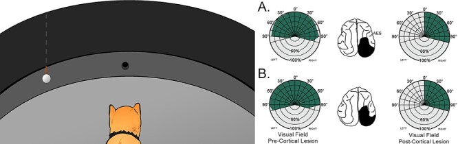
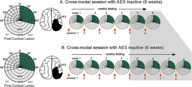
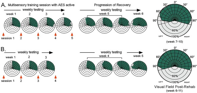
Hemianopia induced by unilateral visual cortex lesions can be resolved by repeatedly exposing the blinded hemifield to auditory-visual stimuli. This rehabilitative "training" paradigm depends on mechanisms of multisensory plasticity that restore the lost visual responsiveness of multisensory neurons in the ipsilesional superior colliculus (SC) so that they can once again support vision in the blinded hemifield. These changes are thought to operate via the convergent visual and auditory signals relayed to the SC from association cortex (the anterior ectosylvian sulcus [AES], in cat). The present study tested this assumption by cryogenically deactivating ipsilesional AES in hemianopic, anesthetized cats during weekly multisensory training sessions. No signs of visual recovery were evident in this condition, even after providing animals with up to twice the number of training sessions required for effective rehabilitation. Subsequent training under the same conditions, but with AES active, reversed the hemianopia within the normal timeframe. These results indicate that the corticotectal circuit that is normally engaged in SC multisensory plasticity has to be operational for the brain to use visual-auditory experience to resolve hemianopia.

Keywords: colliculus; ectosylvian; hemianopia; multisensory; rehabilitation.


Figures

Figure 1
Figure 1
The visual localization task. Left: Animals were first trained to fixate a food reward at 0°, and then to approach a ping pong ball lowered from behind an opaque curtain in 15° (randomly presented) steps of eccentricity from 0° to ±105°. Little training was required to master this task, and each animal responded with high reliability to the stimulus at all locations. Right: Visual detection and orientation performance before (pre-) and after (post-) the hemianopia-inducing lesion in 2 animals (A, B) implanted with cryogenic coils in AES. Each circle in the polar plot represents 20% correct performance (shown in green), and each radial line = 15° of eccentricity. Schematics of the brain illustrate cortical lesion in black. Note the absence of contralesional visual responses after the lesion.
Figure 2
Figure 2
Multisensory training with AES deactivated was ineffective in resolving hemianopia. Hemianopic animals given 6 (A) or 8 (B) weeks of training with spatiotemporally congruent visual–auditory stimuli in the blinded hemifield (left) while the ipsilesional (right) AES was deactivated. On the left are radial plots showing visual localization performance prior to rehabilitative training. On the right are radial plots of visual performance in which data were averaged over each week of rehabilitative training. Note the absence of visual recovery. Shaded region indicates testing points at which the control animals had already recovered visual responsiveness. Other conventions are the same as in Fig 1.
Figure 3
Figure 3
Subsequent multisensory training without AES deactivation resolved hemianopia. The same training that was previously ineffective when AES was deactivated now rehabilitated hemianopia within the normal time period in both animals (A, B). Visual responses first appeared at central locations within 4 weeks of training onset, and then progressed peripherally without further training. By week 6 vision had been restored throughout the contralesional hemifield and remained stable thereafter. Conventions are the same as previous figures.

Similar articles

Cited by

References

    1. Alvarado JC, Stanford TR, Rowland BA, Vaughan JW, Stein BE. 2009. Multisensory integration in the superior colliculus requires synergy among corticocollicular inputs. J Neurosci. 29(20):6580–6592. doi: 10.1523/JNEUROSCI.0525-09.2009. - DOI - PMC - PubMed
    1. Alvarado JC, Stanford TR, Vaughan JW, Stein BE. 2007. Cortex mediates multisensory but not unisensory integration in superior colliculus. J Neurosci. 27(47):12775–12786. doi: 10.1523/JNEUROSCI.3524-07.2007. - DOI - PMC - PubMed
    1. Benedek G, Keri S, Nagy A, Braunitzer G, Norita M. 2019. A multimodal pathway including the basal ganglia in the feline brain. Physiol Int. 106(2):95–113. doi: 10.1556/2060.106.2019.09. - DOI - PubMed
    1. Benedek G, Perény J, Kovács G, Fischer-Szátmári L, Katoh YY. 1997. Visual, somatosensory, auditory and nociceptive modality properties in the feline suprageniculate nucleus. Neuroscience. 78(1):179–189. doi: 10.1016/s0306-4522(96)00562-3. - DOI - PubMed
    1. Brzosko Z, Mierau SB, Paulsen O. 2019. Neuromodulation of spike-timing-dependent plasticity: past, present, and future. Neuron. 103(4):563–581. doi: 10.1016/j.neuron.2019.05.041. - DOI - PubMed

Publication types