. 2021 May 31;122(9):1021-1036.
doi: 10.1002/jcb.29907. Online ahead of print.

Unsulfated biotechnological chondroitin by itself as well as in combination with high molecular weight hyaluronan improves the inflammation profile in osteoarthritis in vitro model

Affiliations

Unsulfated biotechnological chondroitin by itself as well as in combination with high molecular weight hyaluronan improves the inflammation profile in osteoarthritis in vitro model

Valentina Vassallo et al. J Cell Biochem. .

Abstract

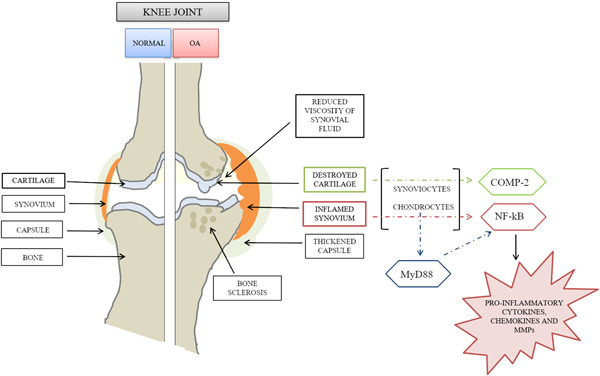
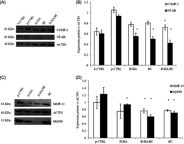
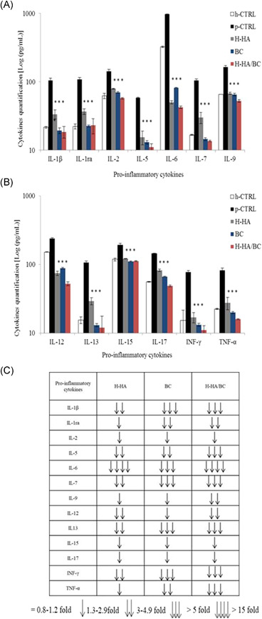
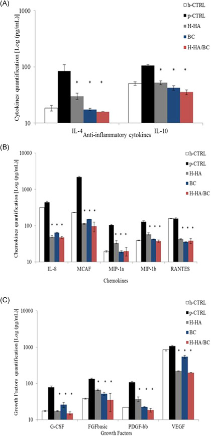
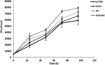
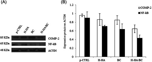
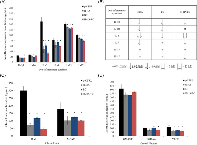
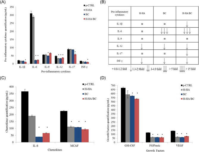
Several studies suggest that inflammation has a pivotal role during the progression of osteoarthritis (OA) and cytokines have been identified as the main process mediators. This study aimed to explore the ability to modulate the main OA pro-inflammatory biomarkers of novel gels (H-HA/BC) based on high molecular weight hyaluronan (H-HA) and unsulfated biotechnological chondroitin (BC). For the first time, BC was tested also in combination with H-HA on human primary cells isolated from pathological knee joints. Specifically, the experiments were performed using an OA in vitro model based on human chondrocytes and synoviocytes. To evaluate the anti-inflammatory effects of H-HA/BC in comparison with H-HA and BC single gels, NF-kB, COMP-2, MyD88, MMP-13 and a wide range of cytokines, known to be specific biomarkers in OA (e.g., IL-6, IL-8, and TNF-α), were evaluated. In addition, cell morphology and proliferation occurring in the presence of either H-HA/BC or single components were assessed using time-lapse video microscopy. It was shown that synovial fluids and cells isolated from OA suffering patients, presented a cytokine pattern respondent to an ongoing inflammation status. H-HA and BC significantly reduced the levels of 23 biomarkers associated with cartilage damage. However, H-HA/BC decreased significantly 24 biological mediators and downregulated 19 of them more efficiently than the single components. In synoviocytes cultures, cytokine analyses proved that H-HA/BC gels re-established an extracellular environment more similar to a healthy condition reducing considerably the concentration of 11 analytes. Instead, H-HA and BC significantly modulated 7 (5 only with a longer treatment) and 8 biological cytokines, respectively. Our results suggest that H-HA/BC beyond the viscosupplementation effect typical for HA-based gels, can improve the inflammation status in joints and thus could be introduced as a valid protective and anti-inflammatory intraarticular device in the field of Class III medical devices for OA treatments.

Keywords: biotechnological chondroitin; human articular chondrocytes; human synoviocytes; hybrid cooperative complexes; inflammation; osteoarthritis.


Conflict of interest statement

The authors declare that there are no conflict of interests.

Figures

Figure 1
Figure 1
Schematic representation of the explored degenerative joint process and highlight of the selected biomarkers analyzed
Figure 2
Figure 2
Histological analyses: Microscopic evaluation of Safranin‐O/Fast Green Staining. Histologic sections were observed under light microscopy at low magnification: ×4 (A and C) and ×10 (B and D). Panel A and B are representative of less damaged tissue while C and D represent a more compromised sampling area
Figure 3
Figure 3
Western blot analysis: evaluation of COMP‐2, NF‐kB, MMP‐13, and MyD88 protein levels in primary articular chondrocytes treated for 48 h with H‐HA, BC, and H‐HA/CB 0.32% w/v. (A) Specifics bands obtained for COMP‐2, NF‐kB, and ACTIN. (B) Densitometric analysis performed normalizing COMP‐2 and NF‐kB protein expression with respect to ACTIN. (C) One of the western blots run is reported for MMP‐13, MyD88, and ACTIN. (D) Densitometric analysis was performed for two different western blot experiments normalizing MMP‐13 and MyD88 protein expression with respect to ACTIN. *p <  0.05 t‐test analyses were performed to compare the significance of each treatment with respect to p‐CTRL. BC, biotechnological chondroitin; COMP‐2, cartilage oligomeric matrix protein 2; H‐HA, high molecular weight hyaluronan; h‐CTRL, healthy control; p‐CTRL, pathological control; MyD88, cytosolic adaptor myeloid differentiation factor 88; MMP‐13, matrix metalloproteases; NF‐kB, nuclear factor kappa‐light‐chain‐enhancer of activated B cells
Figure 4
Figure 4
Bio‐Plex assay: Evaluation of biological mediator modulation in supernatants (pg/ml), of chondrocytes treated for 48 h with H‐HA, BC, or H‐HA/BC 0.32% w/v. (A) and (B) Pro‐inflammatory cytokines, (C) the table shows the pro‐inflammatory cytokine negative modulations of at least 30% with respect to p‐CTRL for each treatment. *p <  0.05 t‐test analyses were performed to compare the significance of each treatment with respect to p‐CTRL. BC, biotechnological chondroitin; H‐HA, high molecular weight hyaluronan; h‐CTRL, healthy control; IL, interleukin; INF, interferon; p‐CTRL, pathological control; TNF‐α: Tumor necrosis factor α
Figure 5
Figure 5
Bio‐Plex assay: (A) Anti‐inflammatory cytokines, (B) chemokines, (C) growth factors. *p < .05 t‐test analyses were performed to compare the significance of each treatment with respect to p‐CTRL. BC, biotechnological chondroitin; H‐HA, high molecular weight hyaluronan; h‐CTRL, healthy control; IL, interleukin; p‐CTRL, pathological control; MCAF, monocyte chemotactic and activating factor
Figure 6
Figure 6
Human synoviocytes proliferation assay H‐HA/BC were compared with the single H‐HA, BC, and no treated cells (p‐CTRL) on cell proliferation. Cells growth curves were obtained through Image‐Pro Plus 1.5 software (Media Cybernetics). BC, biotechnological chondroitin; H‐HA, high molecular weight hyaluronan; p‐CTRL, pathological control
Figure 7
Figure 7
Western blot analysis evaluation of COMP‐2 and NF‐kB protein levels in synoviocytes treated for 48 h with H‐HA, BC, and H‐HA/CB 0.32% w/v. (A) Specific bands obtained for COMP‐2, NF‐kB, and ACTIN. (B) Densitometric analysis performed normalizing COMP‐2 and NF‐kB protein expression with respect to ACTIN. *p <  0.05 t‐test analyses were performed to compare the significance of each treatment with respect to p‐CTRL. BC, biotechnological chondroitin; COMP‐2, cartilage oligomeric matrix protein 2; H‐HA, high molecular weight hyaluronan; p‐CTRL, pathological control; NF‐kB, nuclear factor kappa‐light‐chain‐enhancer of activated B cells
Figure 8
Figure 8
Bio‐Plex assay: evaluation of biological mediators modulation in supernatants (pg/ml), of synoviocytes treated for 8 h with H‐HA, BC, or H‐HA/BC 0.32% w/v in free serum culture media. (A) Pro‐inflammatory cytokines, (B) the table shows the pro‐inflammatory cytokine negative modulations of at least 30% with respect to p‐CTRL for each treatment, (C) chemokines, and (D) growth factors. *p <  0.05 t‐test analyses were performed to compare the significance of each treatment with respect to p‐CTRL. BC, biotechnological chondroitin; H‐HA, high molecular weight hyaluronan; IL, interleukin; p‐CTRL, pathological control; MCAF, monocyte chemotactic and activating factor
Figure 9
Figure 9
Bio‐Plex assay: evaluation of biological mediators modulation in supernatants (pg/ml), of synoviocytes treated for 48 h with H‐HA, BC, or H‐HA/BC 0.32% w/v in free serum culture media. (A) Pro‐inflammatory cytokines, (B) the table shows the pro‐inflammatory cytokine negative modulations of at least 30% with respect to p‐CTRL for each treatment, (C) chemokines, and (D) growth factors. *p <  0.05 t‐test analyses were performed to compare the significance of each treatment with respect to p‐CTRL. BC, biotechnological chondroitin; H‐HA, high molecular weight hyaluronan; IL, interleukin; p‐CTRL, pathological control; MCAF, monocyte chemotactic and activating factor

Similar articles

Cited by

References

    1. Ueda K, Sasaki N, Goren A, et al. Treatment satisfaction with pharmaceutical interventions in Japanese adults with osteoarthritis and chronic knee pain: an analysis of a web‐based survey. Clin Interv Aging. 2018;13:2179‐2191. 10.2147/CIA.S176289 - DOI - PMC - PubMed
    1. Johnson CI, Argyle D, Clements DN. In vitro models for the study of osteoarthritis. Vet J. 2016;209:40‐49. 10.1016/j.tvjl.2015.07.011 - DOI - PubMed
    1. Loeser RF, Goldring SR, Scanzello CR, Goldring MB. Osteoarthritis: a disease of the joint as an organ. Arthritis Rheum. 2012;64(6):1697‐1707. 10.1002/art.34453 - DOI - PMC - PubMed
    1. Calamia V, Fernàndez‐Puente P, Mateos J, et al. Pharmacoproteomic study of three different chondroitin sulfate compounds on intracellular and extracellular human choncrocyte proteomes. Mol Cell Proteomics. 2012;11(6):M111.013417 - PMC - PubMed
    1. Hochberg MC, Altman RD, April KT, et al. American College of Rheumatology 2012 recommendations for the use of non pharmacologic and pharmacologic therapies in osteoarthritis of the hand, hip, and knee. Arthritis Care Res (Hoboken). 2012;4:465‐474. - PubMed