Review
. 2022 Feb;111(2):497-508.
doi: 10.1002/JLB.4COVHR0221-093RR. Epub 2021 May 31.

Inflammasome regulation in driving COVID-19 severity in humans and immune tolerance in bats

Affiliations
Review

Inflammasome regulation in driving COVID-19 severity in humans and immune tolerance in bats

Sahana Nagaraja et al. J Leukoc Biol. 2022 Feb.

Abstract

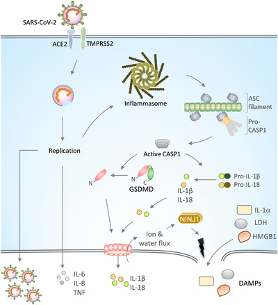
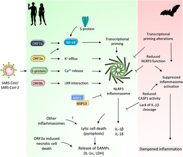
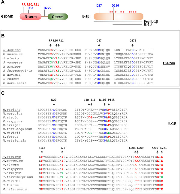
Coronaviruses (CoVs) are RNA viruses that cause human respiratory infections. Zoonotic transmission of the SARS-CoV-2 virus caused the recent COVID-19 pandemic, which led to over 2 million deaths worldwide. Elevated inflammatory responses and cytotoxicity in the lungs are associated with COVID-19 severity in SARS-CoV-2-infected individuals. Bats, which host pathogenic CoVs, operate dampened inflammatory responses and show tolerance to these viruses with mild clinical symptoms. Delineating the mechanisms governing these host-specific inflammatory responses is essential to understand host-virus interactions determining the outcome of pathogenic CoV infections. Here, we describe the essential role of inflammasome activation in determining COVID-19 severity in humans and innate immune tolerance in bats that host several pathogenic CoVs. We further discuss mechanisms leading to inflammasome activation in human SARS-CoV-2 infection and how bats are molecularly adapted to suppress these inflammasome responses. We also report an analysis of functionally important residues of inflammasome components that provide new clues of bat strategies to suppress inflammasome signaling and innate immune responses. As spillover of bat viruses may cause the emergence of new human disease outbreaks, the inflammasome regulation in bats and humans likely provides specific strategies to combat the pathogenic CoV infections.

Keywords: Bat immunity; COVID-19; Coronaviruses; Immune tolerance; Inflammasome; NLRP3; SARS-CoV-2.


Figures

FIGURE 1
FIGURE 1
SARS‐CoV‐2 infection, inflammasome activation, and release of proinflammatory cytokines. Spike (S) protein of the SARS‐CoV‐2 binds to the ACE2 receptor on the host cell membrane for its entry into the cell. The cell surface protease TMPRSS2 cleaves S protein to enable membrane fusion and entry of the virus. The replication of the viral RNA genome generates new SARS‐CoV‐2 virions. SARS‐CoV‐2 replication triggers the activation and assembly of the inflammasome and release of IL‐1α, IL‐1β, and IL‐18 cytokines that are implicated in COVID‐19 severity. Upon inflammasome activation, ASC oligomerizes into large filamentous structures/speck‐like structures that recruit and activate CASP1 via CARD‐mediated homotypic interactions. Active CASP1 cleaves pro‐IL‐1β and pro‐IL‐18 into mature forms. The CASP1 also cleaves GSDMD to liberate its N‐terminal domain that oligomerizes into a pore‐structure on the plasma membrane. GSDMD pores facilitate the passage of ions, water, and small molecules like IL‐1β and IL‐18. GSDMD pore formation licenses plasma membrane rupture through NINJ1 and triggers lytic cell death. Plasma membrane rupture is crucial for releasing large molecular weight DAMPs like IL‐1α, LDH, and HMGB1. Inflammasome‐independent cytokines, IL‐6, IL‐8, and TNF, secreted in response to SARS‐CoV‐2 infection, are associated with CoVID‐19 severity
FIGURE 2
FIGURE 2
Differential regulation of inflammasome activation in bats and humans and its impact on CoV‐induced immune responses. Pathogenic CoVs (SARS‐CoV and SARS‐CoV‐2) trigger activation of the NLRP3 inflammasome in host cells. SARS‐CoV‐2 viroporins, ORF3a and E protein promote potassium (K+) efflux or calcium (Ca2+) release into the cytosol, which activates the NLRP3 inflammasome. These viroporins also stimulate NF‐kB activation and transcriptional priming of NLRP3, IL‐1b and IL‐18. ORF7a and S proteins also promote priming and NF‐kB activation. ORF8b of the SARS‐CoV interacts with the LRR domain of the NLRP3 to promote its activation. The ORF3a and S protein of SARS‐CoV‐2 also engage NLRP3 inflammasome activation. NSP1 and NSP13 proteins of the SARS‐CoV‐2 abolish NLRP3 inflammasome activation and CASP1 mediated IL‐1β release. The inflammasome activation and release of IL‐1β and IL‐18 correlate with COVID‐19 severity in SARS‐CoV‐2 infected patients. Other inflammasomes or lytic cell death routes (ORF3a induced necrotic cell death) may also promote the release of DAMPs and excessive inflammatory responses. Bats have adapted to suppress NLRP3 inflammasome activation through regulating transcriptional priming and assembly of the inflammasome complex. This inflammasome suppression in bats dampens inflammation and confer immune tolerance to pathogenic CoVs
FIGURE 3
FIGURE 3
Bats show variation in functionally essential residues of inflammasome components. (A) Domain architectures of the ASC and CASP1 proteins. Asterisks indicate critical residue positions promoting homotypic interactions and oligomerization of ASC and CASP1; cleavage site residues of CASP1 are indicated in blue; the catalytically important residue of the CASP1 are indicated in red. (B) Common and scientific names of organisms that are used in panels C and D for comparing the conservation of inflammasomes components. Bat species are categorized into Yangochiroptera and Yinpterochiroptera suborders. (C and D) Multiple sequence alignments of specific regions of ASC (C) and CASP1 (D) from human, mouse, and bat species. The human ASC and CASP1 protein sequences are used as references to define the numbers of amino acid residues. The critical residue positions are colored as represented in panel A. The conserved residue positions are highlighted in red, the conserved cleavage site residues are in blue, and the mutated residue positions in green. The D122m in panel D represents the cleavage site in mice that correspond to the D119 cleavage site of the human CASP1
FIGURE 4
FIGURE 4
Variations in functionally important residues of bat CASP1 substrates, GSDMD and IL‐1β proteins. (A) Cartoon representing GSDMD, pro‐IL‐1β and mature form of IL‐1β. Asterisks indicate critical residue positions required for IL‐1β function; cleavage site residues are indicated in blue; critical lipid membrane binding residues of the GSDMD are indicated in red. (B and C) Multiple sequence alignments showing critical residues of GSDMD (B) and IL‐1β protein (C). The human (H.sapiens) GSDMD and IL‐1β protein sequences are used as a reference sequence to represent the amino acid residue numbers. The conserved amino acid residues are highlighted in red. The conserved cleavage site residue positions are highlighted in blue. The mutated amino acid residue positions are highlighted in green

Similar articles

Cited by

References

    1. Cui J, Li F, Shi Z‐Li. Origin and evolution of pathogenic coronaviruses. Nat Rev Microbiol. 2019;17:181‐192. - PMC - PubMed
    1. V'kovski P, Kratzel A, Steiner S, Stalder H, Thiel V. Coronavirus biology and replication: implications for SARS‐CoV‐2. Nat Rev Microbiol. 2020. - PMC - PubMed
    1. Weiss SR. Forty years with coronaviruses. J Exp Med. 2020;217. - PMC - PubMed
    1. Hoffmann M, Kleine‐Weber H, Schroeder S, et al. SARS‐CoV‐2 cell entry depends on ACE2 and TMPRSS2 and is blocked by a clinically proven protease inhibitor. Cell. 2020;181:271‐280 e278. - PMC - PubMed
    1. Lan J, Ge J, Yu J, et al. Structure of the SARS‐CoV‐2 spike receptor‐binding domain bound to the ACE2 receptor. Nature. 2020;581:215‐220. - PubMed

Publication types