

Peters plus syndrome mutations affect the function and stability of human β1,3-glucosyltransferase
- PMID: 34058199
- PMCID: PMC8233153
- DOI: 10.1016/j.jbc.2021.100843
Peters plus syndrome mutations affect the function and stability of human β1,3-glucosyltransferase
Abstract
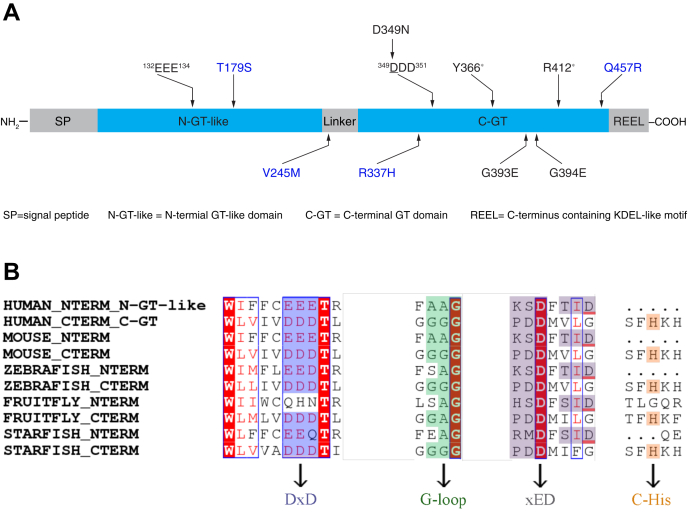
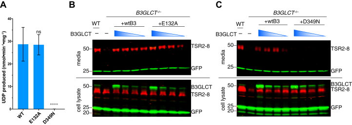
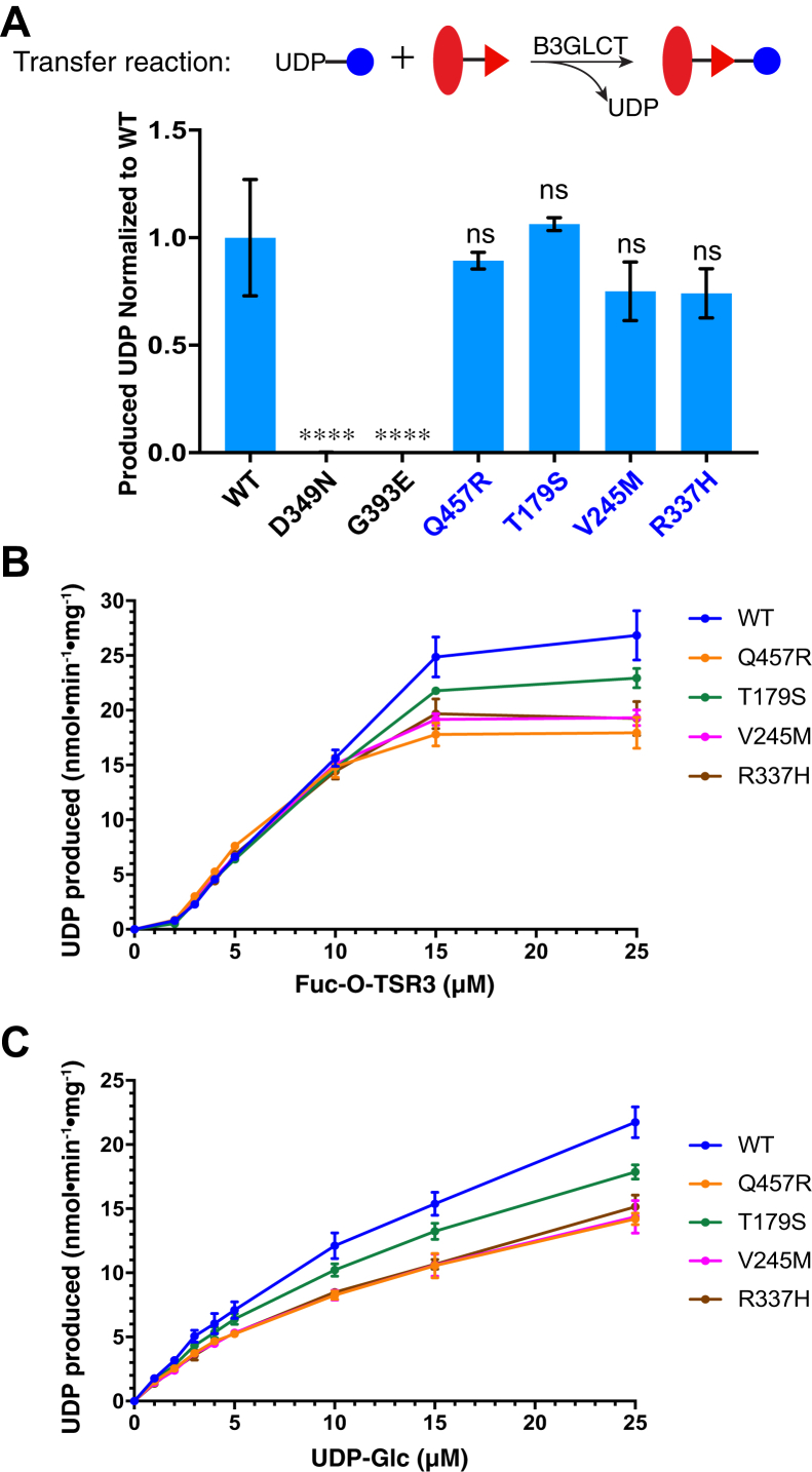
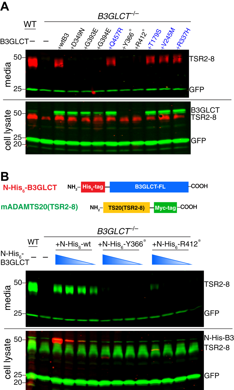
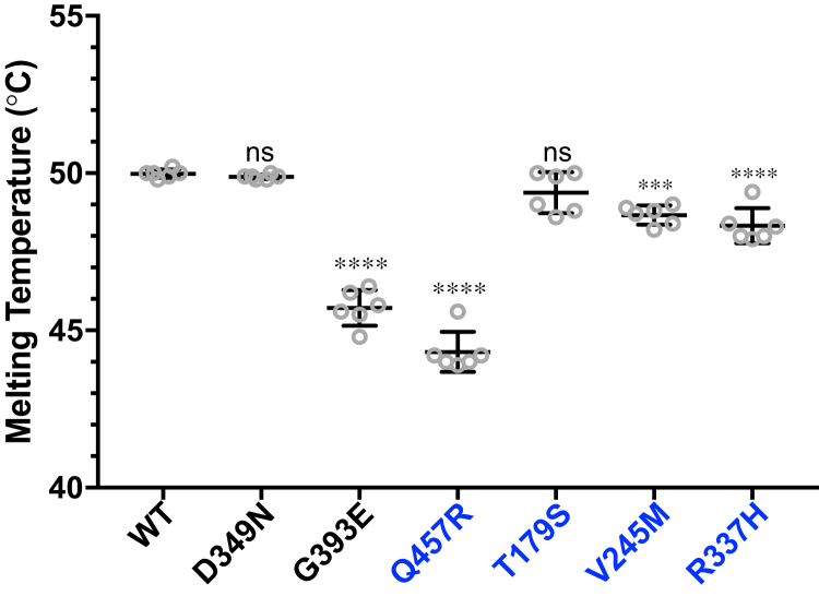
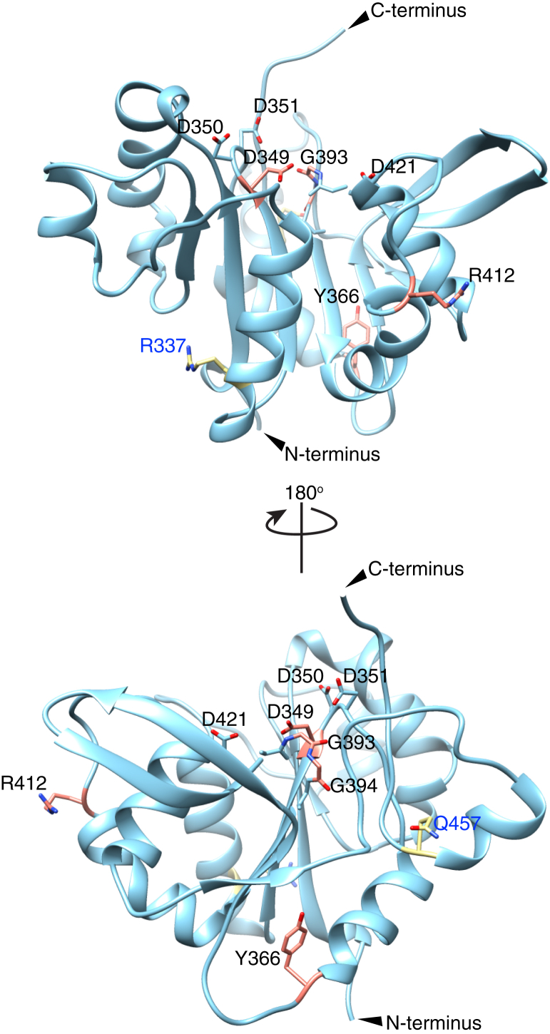
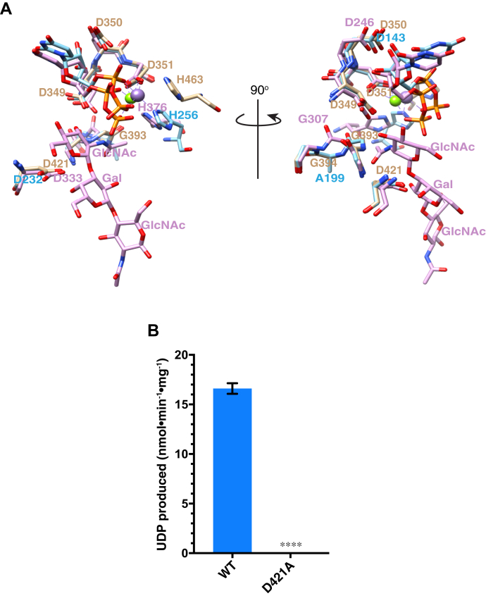
Peters Plus Syndrome (PTRPLS OMIM #261540) is a severe congenital disorder of glycosylation where patients have multiple structural anomalies, including Peters anomaly of the eye (anterior segment dysgenesis), disproportionate short stature, brachydactyly, dysmorphic facial features, developmental delay, and variable additional abnormalities. PTRPLS patients and some Peters Plus-like (PTRPLS-like) patients (who only have a subset of PTRPLS phenotypes) have mutations in the gene encoding β1,3-glucosyltransferase (B3GLCT). B3GLCT catalyzes the transfer of glucose to O-linked fucose on thrombospondin type-1 repeats. Most B3GLCT substrate proteins belong to the ADAMTS superfamily and play critical roles in extracellular matrix. We sought to determine whether the PTRPLS or PTRPLS-like mutations abrogated B3GLCT activity. B3GLCT has two putative active sites, one in the N-terminal region and the other in the C-terminal glycosyltransferase domain. Using sequence analysis and in vitro activity assays, we demonstrated that the C-terminal domain catalyzes transfer of glucose to O-linked fucose. We also generated a homology model of B3GLCT and identified D421 as the catalytic base. PTRPLS and PTRPLS-like mutations were individually introduced into B3GLCT, and the mutated enzymes were evaluated using in vitro enzyme assays and cell-based functional assays. Our results demonstrated that PTRPLS mutations caused loss of B3GLCT enzymatic activity and/or significantly reduced protein stability. In contrast, B3GLCT with PTRPLS-like mutations retained enzymatic activity, although some showed a minor destabilizing effect. Overall, our data supports the hypothesis that loss of glucose from B3GLCT substrate proteins is responsible for the defects observed in PTRPLS patients, but not for those observed in PTRPLS-like patients.
Keywords: B3GLCT; O-fucose; Peters plus syndrome; enzyme catalysis; genetic disease; glycobiology; glycoprotein secretion; glycosyltransferase; thrombospondin type-1 repeats.
Copyright © 2021 The Authors. Published by Elsevier Inc. All rights reserved.
Conflict of interest statement
Conflict of interest The authors declare that they have no conflicts of interest with the contents of this article.
Figures
Similar articles
-
ADAMTS9 and ADAMTS20 are differentially affected by loss of B3GLCT in mouse model of Peters plus syndrome.Hum Mol Genet. 2019 Dec 15;28(24):4053-4066. doi: 10.1093/hmg/ddz225. Hum Mol Genet. 2019. PMID: 31600785 Free PMC article.
-
Peters plus syndrome mutations disrupt a noncanonical ER quality-control mechanism.Curr Biol. 2015 Feb 2;25(3):286-295. doi: 10.1016/j.cub.2014.11.049. Epub 2014 Dec 24. Curr Biol. 2015. PMID: 25544610 Free PMC article.
-
Functional characterization of zebrafish orthologs of the human Beta 3-Glucosyltransferase B3GLCT gene mutated in Peters Plus Syndrome.PLoS One. 2017 Sep 19;12(9):e0184903. doi: 10.1371/journal.pone.0184903. eCollection 2017. PLoS One. 2017. PMID: 28926587 Free PMC article.
-
Ocular Phenotype of Peters-Plus Syndrome.Cornea. 2022 Feb 1;41(2):219-223. doi: 10.1097/ICO.0000000000002889. Cornea. 2022. PMID: 34629439 Review.
-
Peters'-plus syndrome is a congenital disorder of glycosylation caused by a defect in the beta1,3-glucosyltransferase that modifies thrombospondin type 1 repeats.Ann Med. 2009;41(1):2-10. doi: 10.1080/07853890802301975. Ann Med. 2009. PMID: 18720094 Review.
Cited by
-
Congenital disorders of glycosylation (CDG): state of the art in 2022.Orphanet J Rare Dis. 2023 Oct 19;18(1):329. doi: 10.1186/s13023-023-02879-z. Orphanet J Rare Dis. 2023. PMID: 37858231 Free PMC article. Review.
-
Congenital anterior segment ocular disorders: Genotype-phenotype correlations and emerging novel mechanisms.Prog Retin Eye Res. 2024 Sep;102:101288. doi: 10.1016/j.preteyeres.2024.101288. Epub 2024 Aug 2. Prog Retin Eye Res. 2024. PMID: 39097141 Review.
-
Passing the post: roles of posttranslational modifications in the form and function of extracellular matrix.Am J Physiol Cell Physiol. 2023 May 1;324(5):C1179-C1197. doi: 10.1152/ajpcell.00054.2023. Epub 2023 Mar 13. Am J Physiol Cell Physiol. 2023. PMID: 36912485 Free PMC article. Review.
-
O-fucosylation stabilizes the TSR3 motif in thrombospondin-1 by interacting with nearby amino acids and protecting a disulfide bond.J Biol Chem. 2022 Jun;298(6):102047. doi: 10.1016/j.jbc.2022.102047. Epub 2022 May 18. J Biol Chem. 2022. PMID: 35597280 Free PMC article.
-
Glycosylation and behavioral symptoms in neurological disorders.Transl Psychiatry. 2023 May 8;13(1):154. doi: 10.1038/s41398-023-02446-x. Transl Psychiatry. 2023. PMID: 37156804 Free PMC article. Review.
References
-
- Aliferis K., Marsal C., Pelletier V., Doray B., Weiss M.M., Tops C.M., Speeg-Schatz C., Lesnik S.A., Dollfus H. A novel nonsense B3GALTL mutation confirms Peters plus syndrome in a patient with multiple malformations and Peters anomaly. Ophthalmic Genet. 2010;31:205–208. - PubMed
-
- Weh E., Reis L.M., Tyler R.C., Bick D., Rhead W.J., Wallace S., McGregor T.L., Dills S.K., Chao M.C., Murray J.C., Semina E.V. Novel B3GALTL mutations in classic Peters plus syndrome and lack of mutations in a large cohort of patients with similar phenotypes. Clin. Genet. 2014;86:142–148. - PMC - PubMed
-
- Heinonen T.Y., Pasternack L., Lindfors K., Breton C., Gastinel L.N., Maki M., Kainulainen H. A novel human glycosyltransferase: Primary structure and characterization of the gene and transcripts. Biochem. Biophys. Res. Commun. 2003;309:166–174. - PubMed
-
- Sato T., Sato M., Kiyohara K., Sogabe M., Shikanai T., Kikuchi N., Togayachi A., Ishida H., Ito H., Kameyama A., Gotoh M., Narimatsu H. Molecular cloning and characterization of a novel human beta1,3-glucosyltransferase, which is localized at the endoplasmic reticulum and glucosylates O-linked fucosylglycan on thrombospondin type 1 repeat domain. Glycobiology. 2006;16:1194–1206. - PubMed
Publication types
MeSH terms
Substances
Supplementary concepts
Grants and funding
LinkOut - more resources
Full Text Sources
Medical
Molecular Biology Databases
Miscellaneous
