Review
. 2022 Jul;107(7):665-673.
doi: 10.1113/EP089474. Epub 2021 Jun 18.

The role of lactate in sepsis and COVID-19: Perspective from contracting skeletal muscle metabolism

Affiliations
Review

The role of lactate in sepsis and COVID-19: Perspective from contracting skeletal muscle metabolism

Ulrik Winning Iepsen et al. Exp Physiol. 2022 Jul.

Abstract

New findings: What is the topic of this review? Lactate is considered an important substrate for mitochondria in the muscles, heart and brain during exercise and is the main gluconeogenetic precursor in the liver and kidneys. In this light, we review the (patho)physiology of lactate metabolism in sepsis and coronavirus disease 2019 (COVID-19). What advances does it highlight? Elevated blood lactate is strongly associated with mortality in septic patients. Lactate seems unrelated to tissue hypoxia but is likely to reflect mitochondrial dysfunction and high adrenergic stimulation. Patients with severe COVID-19 exhibit near-normal blood lactate, indicating preserved mitochondrial function, despite a systemic hyperinflammatory state similar to sepsis.

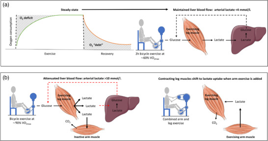
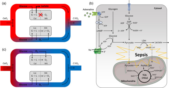
Abstract: In critically ill patients, elevated plasma lactate is often interpreted as a sign of organ hypoperfusion and/or tissue hypoxia. This view on lactate is likely to have been influenced by the pioneering exercise physiologists around 1920. August Krogh identified an oxygen deficit at the onset of exercise that was later related to an oxygen 'debt' and lactate accumulation by A. V. Hill. Lactate is considered to be the main gluconeogenetic precursor in the liver and kidneys during submaximal exercise, but hepatic elimination is attenuated by splanchnic vasoconstriction during high-intensity exercise, causing an exponential increase in blood lactate. With the development of stable isotope tracers, lactate has become established as an important energy source for muscle, brain and heart tissue, where it is used for mitochondrial respiration. Plasma lactate > 4 mM is strongly associated with mortality in septic shock, with no direct link between lactate release and tissue hypoxia. Herein, we provide evidence for mitochondrial dysfunction and adrenergic stimulation as explanations for the sepsis-induced hyperlactataemia. Despite profound hypoxaemia and intense work of breathing, patients with severe coronavirus disease 2019 (COVID-19) rarely exhibit hyperlactataemia (> 2.5 mM), while presenting a systemic hyperinflammatory state much like sepsis. However, lactate dehydrogenase, which controls the formation of lactate, is markedly elevated in plasma and strongly associated with mortality in severe COVID-19. We briefly review the potential mechanisms of the lactate dehydrogenase elevation in COVID-19 and its relationship to lactate metabolism based on mechanisms established in contracting skeletal muscle and the acute respiratory distress syndrome.

Keywords: acute respiratory distress syndrome; cardiovascular system; critical care; exercise; lung injury.


Conflict of interest statement

None declared.

Figures

FIGURE 1
FIGURE 1
Lactate metabolism during steady‐state exercise (a) and incremental exercise (b). (a) The deficit (shaded area, left side) in oxygen consumption (V˙O2) at the onset of exercise was thought to be related to the accumulation of blood lactate, which was degraded during recovery as a waste product of metabolism (i.e., O2 debt). The right side illustrates, however, that arterial lactate is maintained < 4 mmol/l during steady‐state bicycle exercise for 2 h at 60% of maximal V˙O2 (V˙O2max) by gluconeogenesis in the liver and kidneys (not shown). (b) The left side shows that the exponential increase in arterial lactate during incremental exercise is explained, in part, by splanchnic vasoconstriction and thereby reduced gluconeogenesis even though lactate uptake is increased by the inactive arm muscles. Contracting leg muscles are main producers of lactate, but by adding arm muscle exercise to exhaustion, the leg muscles shift to lactate uptake (b, right side). Thus, the accumulation of lactate during incremental exercise does not represent an anaerobic threshold
FIGURE 2
FIGURE 2
Intracellular lactate metabolism in muscle and transport to other organs during exercise. Type II muscle fibres (fast‐twitch) are considered to be producers of lactate, whereas type I (slow‐twitch) fibres mainly consume lactate, and there may be an exchange of lactate within a working muscle bed. Lactate is likely to be the end‐product of glycolysis and transported into the mitochondria or out of the muscle fibre. The lactate produced by contracting muscles is transported via the blood to the heart and the brain (bottom right), where it is used as a substrate for mitochondrial respiration, and to the liver and the kidneys (top right), where it is converted to glucose. Abbreviations: Acetyl CoA, acetyl coenzyme A; Glucose‐6‐P, glucose‐6‐phosphate; LDH, lactate dehydrogenase; TCA, tricarboxylic acid cycle
FIGURE 3
FIGURE 3
Sepsis‐induced hyperlactataemia. (a) Sepsis‐associated mitochondrial dysfunction in an organ that normally converts lactate into energy could result in a functional shunt of lactate across the affected organ and cause hyperlactataemia if the capacity of remaining functional mitochondria is exhausted. The model considers all oxygen‐consuming cells in two equally perfused compartments in the hyperdynamic state of sepsis: one with impaired mitochondrial function (top arm) and one normal (bottom arm), where the top arm is the shunted one. Here, blood oxygen content across the vascular bed remains unchanged because of mitochondrial dysfunction while lactate is still produced in the cytosol by glycolysis. Thus, in sepsis, a lactate shunt can exist without clinical signs of tissue hypoxia (central venous saturation < 0.7 is often used as surrogate for tissue hypoxia) or hypoperfusion. (b) Mitochondrial dysfunction and accelerated aerobic glycolysis through adrenergic stimulation during sepsis will result in an excess of lactate. (c) In the absence of mitochondrial dysfunction, arterial hypoxaemia per se will not induce hyperlactataemia unless the oxygen partial pressure in the microcirculation reaches a critical low level, whereby the diffusion gradient of oxygen is no longer sufficient to maintain mitochondrial electron transport. (Modified from Gattinoni et al., .) Abbreviations: AC, adenylate cyclase; Acetyl CoA, acetyl coenzyme A; CaO2 , arterial oxygen content; Cv¯O2, mixed venous oxygen content; Cyt, cytosol; G, glucose; GDP, guanosine diphosphate; Gs, Gs‐protein‐coupled receptor; GTP, guanosine‐5′‐triphosphate; G‐6‐P, glucose‐6‐phosphate; L, lactate; LDH, lactate dehydrogenase; Mit, mitochondria; TCA, tricarboxylic acid cycle; β2, β2‐adrenergic receptor

References

    1. Bassett, D. R. Jr. (2002). Scientific contributions of A. V. Hill: Exercise physiology pioneer. Journal of Applied Physiology, 93, 1567–1582. - PubMed
    1. Brassard, P. , Zaar, M. , Thaning, P. , Secher, N. H. , & Rosenmeier, J. B. (2016). Sympathetic vasoconstrictor responsiveness of the leg vasculature during experimental endotoxemia and hypoxia in humans. Critical Care Medicine, 44, 755–763. 10.1097/CCM.0000000000001486 - DOI - PubMed
    1. Brealey, D. , Brand, M. , Hargreaves, I. , Heales, S. , Land, J. , Smolenski, R. , Davies, N. A. , Cooper, C. E. , & Singer, M. (2002). Association between mitochondrial dysfunction and severity and outcome of septic shock. Lancet, 360, 219–223. 10.1016/S0140-6736(02)09459-X - DOI - PubMed
    1. Brooks, G. A. (2018). The science and translation of lactate shuttle theory. Cell Metabolism, 27, 757–785. 10.1016/j.cmet.2018.03.008 - DOI - PubMed
    1. Brooks, G. A. , Dubouchaud, H. , Brown, M. , Sicurello, J. P. , & Butz, C. E. (1999). Role of mitochondrial lactate dehydrogenase and lactate oxidation in the intracellular lactate shuttle. Proceedings of the National Academy of Sciences of the United States of America, 96, 1129–1134. 10.1073/pnas.96.3.1129 - DOI - PMC - PubMed

Publication types