Case Reports
. 2021 May 31;14(5):e240684.
doi: 10.1136/bcr-2020-240684.

Management of two-level proximal femoral fractures

Affiliations
Case Reports

Management of two-level proximal femoral fractures

Thom Pauline Maria Johannes Vluggen et al. BMJ Case Rep. .

Abstract

We present the case of an 82-year-old female, who experienced a ground-level fall on the trochanter of the right femur. X-rays showed a proximal femoral fracture (PFF) with an unclear and unusual fracture pattern. Three-dimensional CT images were obtained and showed a displaced femoral neck fracture and ipsilateral fracture of the greater trochanter. Our patient underwent unipolar hemiarthroplasty and fixation of the greater trochanter with a hook plate and cable grip. At 11 months, functional outcomes, patient satisfaction and quality of life were excellent. Primary osteoporosis was diagnosed and treatment with bisphosphonates was initiated.Two-level PFFs are rare and complex. Due to ageing and a subsequent increase in osteoporosis, numbers of PFFs with complex fracture patterns might increase in the future. Adequate treatment and early prevention of osteoporosis are key to reduce this risk and lower the overall burden. Surgical treatment should be patient-tailored and focus on minimising the risk of complications and reinterventions.

Keywords: hip implants; hip prosthesis implantation; orthopaedic and trauma surgery; surgery.


Conflict of interest statement

Competing interests: None declared.

Figures

Figure 1
Figure 1
(A) X-ray (anteroposterior view) of the pelvis shows an unusual fracture pattern of the right proximal femur. The fractured greater trochanter is displaced, and the proximal fragment is held in abduction. Additionally, there appears a lytic lesion at the base of the femoral neck with a sclerotic margin. (B) X-ray (axial view) of the right hip shows posterior displacement of the greater trochanteric fracture. Additionally, the femoral neck seems fractured and posteriorly displaced. Overprojection of fragments makes further assessment difficult and a pathological fracture cannot be ruled out.
Figure 2
Figure 2
(A) Ventral view; (B) lateral view; (C) dorsal view. CT scan three-dimensional reconstruction of the pelvis showing a right displaced medial femoral neck fracture (Garden type IV) with comminution. Additionally, there is an ipsilateral greater trochanteric fracture with displacement to cranial and dorsal. The greater trochanteric fragment is held in abduction and external rotation. There are no signs of a pathological fracture.
Figure 3
Figure 3
(A) Exposure of a two-level proximal femoral fracture (displaced greater trochanter; displaced femoral head and neck; and femoral shaft) after posterolateral approach to the hip joint; (B) zoomed view of the displaced femoral head and neck. There is a comminuted fracture zone of the femoral neck and the bone quality appears poor (this seems to be indicative of osteoporosis); (C) exposure after removal of the femoral head and neck. The greater trochanteric fracture is displaced from the femoral shaft. ER, external rotators; FH, femoral head; FN, femoral neck; FS, femoral shaft; GMA, gluteus maximus; GME, gluteus medius; GT, greater trochanter.
Figure 4
Figure 4
(A) Pelvis (anteroposterior view) and (B) hip (axial view) X-ray showing a good position of the unipolar prothesis and an anatomical reduction of the greater trochanteric fracture. There were no signs of complications.
Figure 5
Figure 5
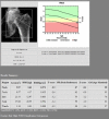
Bone mineral density test indicating osteoporosis of the left hip (total T-score −3.2). ACF, autocorrelation function; BCF, bias correction factor; BMC, bone mineral content; BMD, bone mineral density; CV, coefficient of variation; TH, total hip.
Figure 6
Figure 6
Flowchart for the surgical treatment of proximal femoral fractures. HA, hemiarthroplasty; IF, internal fixation; ORIF, open reduction internal fixation; SDM, shared decision making;THA, total hip arthroplasty.
Figure 7
Figure 7
Flowchart, step-by-step approach to aid the surgical decision of two-level proximal femoral fractures. HA, hemiarthroplasty; IF, internal fixation; PFF, proximal femoral fracture; THA, total hip arthroplasty.

References

    1. Sözen T, Özışık L, Başaran Nursel Çalık, Calik Basaran N. An overview and management of osteoporosis. Eur J Rheumatol 2017;4:46–56. 10.5152/eurjrheum.2016.048 - DOI - PMC - PubMed
    1. Dhanwal DK, Dennison EM, Harvey NC, et al. . Epidemiology of hip fracture: worldwide geographic variation. Indian J Orthop 2011;45:15–22. 10.4103/0019-5413.73656 - DOI - PMC - PubMed
    1. Filipov O. Epidemiology and social burden of the femoral neck fractures. JofIMAB 2014;20:516–8. 10.5272/jimab.2014204.516 - DOI
    1. An HS, Wojcieszek JM, Cooke RF, et al. . Simultaneous ipsilateral intertrochanteric and subcapital fracture of the hip. A case report. Orthopedics 1989;12:721–3. - PubMed
    1. Pemberton DJ, Kriebich DN, Moran CG. Segmental fracture of the neck of the femur. Injury 1989;20:306–7. 10.1016/0020-1383(89)90179-4 - DOI - PubMed

Publication types