. 2021 May 31;6(1):213.
doi: 10.1038/s41392-021-00634-z.

A core-shell structured COVID-19 mRNA vaccine with favorable biodistribution pattern and promising immunity

Affiliations

A core-shell structured COVID-19 mRNA vaccine with favorable biodistribution pattern and promising immunity

Ren Yang et al. Signal Transduct Target Ther. .

Abstract

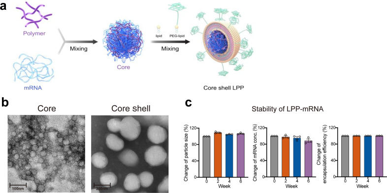
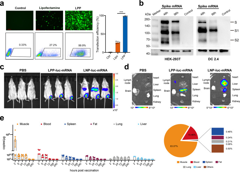
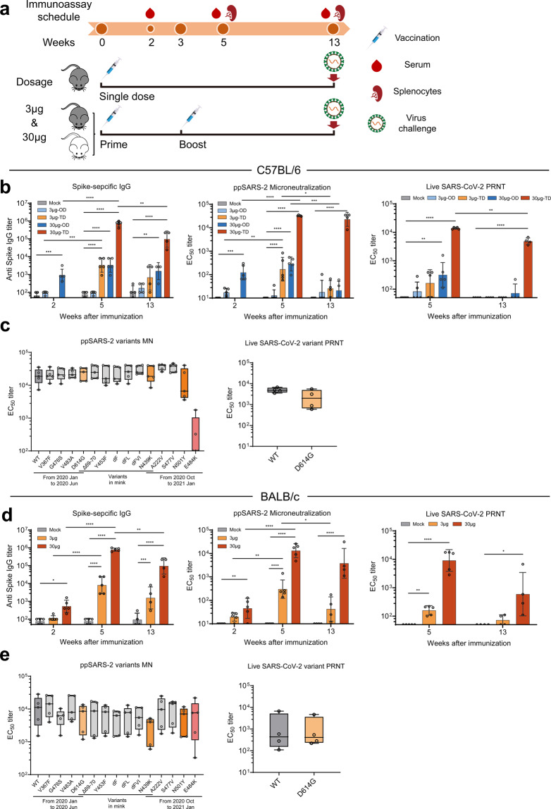
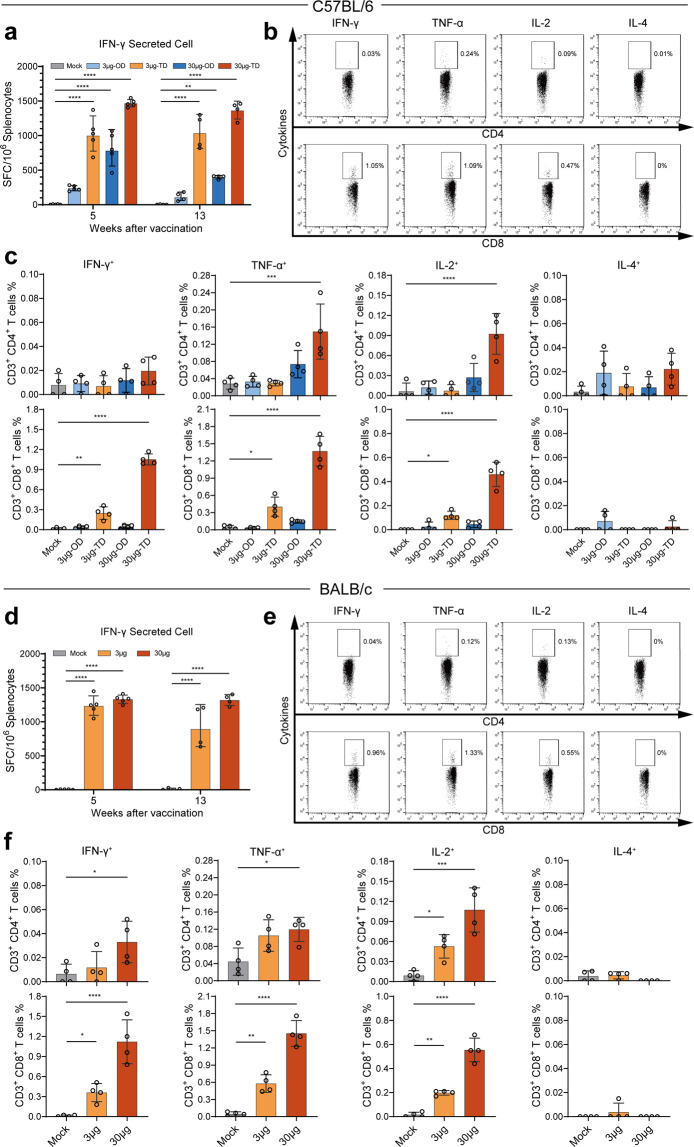
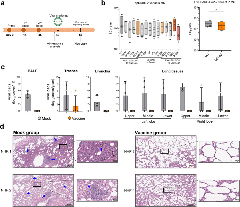
Although inoculation of COVID-19 vaccines has rolled out globally, there is still a critical need for safe and effective vaccines to ensure fair and equitable supply for all countries. Here, we report on the development of a highly efficacious mRNA vaccine, SW0123 that is composed of sequence-modified mRNA encoding the full-length SARS-CoV-2 Spike protein packaged in core-shell structured lipopolyplex (LPP) nanoparticles. SW0123 is easy to produce using a large-scale microfluidics-based apparatus. The unique core-shell structured nanoparticle facilitates vaccine uptake and demonstrates a high colloidal stability, and a desirable biodistribution pattern with low liver targeting effect upon intramuscular administration. Extensive evaluations in mice and nonhuman primates revealed strong immunogenicity of SW0123, represented by induction of Th1-polarized T cell responses and high levels of antibodies that were capable of neutralizing not only the wild-type SARS-CoV-2, but also a panel of variants including D614G and N501Y variants. In addition, SW0123 conferred effective protection in both mice and non-human primates upon SARS-CoV-2 challenge. Taken together, SW0123 is a promising vaccine candidate that holds prospects for further evaluation in humans.


Conflict of interest statement

Stemirna Therapeutics filed patents on patent application entitled “A prophylactic or therapeutical 2019-nCoV mRNA vaccine”. All other authors declare no conflict of interest.

Figures

Fig. 1
Fig. 1
Preparation and characterization of a “core–shell”-structured LPP-mRNA vaccine. a Schematic view on preparation of SW0123 mRNA vaccine. b Representative transmission electronic microscopy (TEM) image of SW0123 vaccine particles. Left: mRNA/SW-01 complexes. Right: LPP particles with a complex lipid cover. c Time-dependent changes in particle size, mRNA concentration and encapsulation efficiency of SW0123 upon storage at 4 °C
Fig. 2
Fig. 2
Transfection efficiency and biodistribution of LPP-mRNA. a eGFP-mRNA was transfected into DC2.4 cells using LPP or lipofectamine. Cells were harvested after 24 h of incubation. eGFP expression was visualized using fluorescent microscope (upper panel) and quantified using flow cytometry (lower panel). b S protein expression from mRNA used for SW0123 preparation. mRNA encoding the S protein was transfected into HEK-293T and DC2.4 immortalized cells. Cells were harvested 48 and 96 h later, and S protein levels in cell lysate were detected with western blot. c Comparison of biodistribution between LPP/mRNA and LNP/mRNA. BALB/c mice were administrated intramuscularly (i.m.) with LPP or LNP encapsulated with luciferase-mRNA. Bioluminescence was measured 6 h later in a Xenogen IVIS-200 imaging system. d Mice were euthanized, and bioluminescent intensity in major organs were detected with a Xenogen IVIS-200 imaging system. e Biodistribution of SW0123 in BALB/c mice. BALB/c mice were i.m. injected with SW0123 at a single dose of 1.5 mg/kg. Major organs or tissues were collected at different time points following vaccination (n = 6 mice each time point). mRNA concentration was determined with real-time qPCR using probes specific for the mRNA component of SW0123 (left panel). Area under curve (AUC) representing accumulative distribution of SW0123 in each specimen is shown (right panel)
Fig. 3
Fig. 3
Induction of robust and persistent levels of Abs with broadly neutralizing capacity by SW0123. a Two different strains of mice were used to evaluate immunogenicity and protective efficacy of vaccines. C57BL/6 mice received either an one-dose (OD) vaccination with 3 or 30 μg SW0123 (based on mRNA content) in week 0 or two-dose (TD) vaccinations with the same dosage in weeks 0 and 3. BALB/c mice were immunized twice with 3 or 30 μg SW0123 in weeks 0 and 3. Mice in the mock group were administrated with empty vector as control. S protein-specific IgG titers were measured in weeks 2, 5, and 13 using ELISA. (n = 5 in weeks 2 and 5; n = 4 in week 13) (b, d left panel). Neutralizing Ab (NAb) titers were measured using a SARS-CoV-2 pseudovirus microneutralization assay (b, d mid panel) or using a live SARS-CoV-2 (Wuhan/IVDC-HB-01/2019) plaque reduction neutralizaion test (b, d right panel). NAb titers are shown as EC50 values, calculated by Reed-Muench method. c, e Serum collected at week 5 from two-dose SW0123-immunized mice (n = 5 for each strain) were tested for neutralizing ability against pseudotype SARS-CoV-2 with indicated mutation points in S protein or were tested against live D614G mutant and wide-type strain (Wuhan/IVDC-HB-01/2019) using plaque reduction neutralization test. Two-tailed Mann–Whitney test was used for statistical analysis. *p ≤ 0.05; **p < 0.01; ***p < 0.001; ****p < 0.0001
Fig. 4
Fig. 4
Induction of predominantly Th1-biased T cell responses by SW0123. C57BL/6 mice received either an one-dose (OD) vaccination with 3 or 30 μg SW0123 in week 0 or two-dose (TD) vaccinations with the same dosage in weeks 0 and 3. BALB/c mice were immunized twice with 3 or 30 μg SW0123 in weeks 0 and 3. Mice in the mock group were administrated with empty vector as control. (n = 5 in week 5; n = 4 in week 13). a, d Splenocytes were isolated 5 and 13 weeks after the first vaccination. Following stimulation with S protein overlapping peptides for 20 h, IFN-γ-producing T cells were quantified with an ELISpot assay. b, c, e, f Splenocytes isolated 13 weeks after first vaccination were stimulated with S protein overlapping peptides for 6 h, and Spike specific cytokine-producing CD4+ and CD8+ T cells were measured with an intracellular cytokine recall assay. Two-tailed Mann–Whitney test was used for statistical analysis. *p ≤ 0.05; **p < 0.01; ***p < 0.001; ****p < 0.0001
Fig. 5
Fig. 5
Induction of efficient protection against SARS-CoV-2 in mice by SW0123. C57BL/6 mice received either an one-dose (OD) vaccination with 3 or 30 μg SW0123 in week 0, or two-dose (TD) vaccinations with the same dosage in weeks 0 and 3. BALB/c mice were immunized twice with 3 or 30 μg SW0123 in weeks 0 and 3. Mice in the mock group were administrated with empty vector as control. Mice (n = 6 each group) were transduced with rAdV5-expressing hACE2 13 weeks after first dosing, and were challenged intranasally with 5 × 105 TCID50 of SARS-CoV-2 (Wuhan/IVDC-HB-01/2019) 5 days of post-transduction. Lung tissues were harvested 4 days of post-challenge. a, c Viral titer levels in the lungs. Right lungs were homogenized and determined for viral titers. b, d Histology of lung sections. Left lungs were sectioned for hematoxylin and eosin (H&E) staining. Two-tailed Mann–Whitney test was used for statistical analysis. ***p < 0.001; ****p < 0.0001
Fig. 6
Fig. 6
Induction of sufficient protection against SARS-CoV-2 in rhesus macaques by SW0123. a Schematic study design. Rhesus macaques received three doses of SW0123, and were intranasally and intratracheally challenged with 1 × 10⁶ PFU of SARS-CoV-2 two weeks after the 2nd boost. Animals in mock group received PBS injection. Samples were collected at the indicated time points. b Titers of NAbs for neutralizing ability against pseudotype SARS-CoV-2 with indicated mutation points in S protein or were tested against live D614G mutant and wide-type strain (Wuhan/IVDC-HB-01/2019) using plaque reduction neutralization test. c Animals were euthanized and necropsied at 7 dpi. gRNA copies in trachea, bronchia, BAL fluid and lung tissues were determined. d H&E staining of lung tissue sections from animals. Blue arrows point to lung pathological lesions. Images from two representative animals in each group are shown. Two-tailed Mann–Whitney test was used for statistical analysis. *p ≤ 0.05; **p < 0.01; ***p < 0.001; ****p < 0.0001

Similar articles

Cited by

References

    1. Thanh Le,T, et al. The COVID-19 vaccine development landscape. Nat. Rev. Drug Discov. 2020;19:305–306. doi: 10.1038/d41573-020-00073-5. - DOI - PubMed
    1. Yu J, et al. DNA vaccine protection against SARS-CoV-2 in rhesus macaques. Science. 2020;369:806–811. doi: 10.1126/science.abc6284. - DOI - PMC - PubMed
    1. Gao Q, et al. Development of an inactivated vaccine candidate for SARS-CoV-2. Science. 2020;369:77–81. doi: 10.1126/science.abc1932. - DOI - PMC - PubMed
    1. Zhu FC, et al. Safety, tolerability, and immunogenicity of a recombinant adenovirus type-5 vectored COVID-19 vaccine: a dose-escalation, open-label, non-randomised, first-in-human trial. Lancet. 2020;395:1845–1854. doi: 10.1016/S0140-6736(20)31208-3. - DOI - PMC - PubMed
    1. Mulligan MJ, et al. Phase 1/2 study to describe the safety and immunogenicity of a COVID-19 RNA vaccine candidate (BNT162b1) in adults 18 to 55 years of age: interim report. Nature. 2020;586:589–593. doi: 10.1038/s41586-020-2639-4. - DOI - PubMed

Publication types

MeSH terms