. 2021 May 31;4(1):645.
doi: 10.1038/s42003-021-02171-5.

WDR62 is required for centriole duplication in spermatogenesis and manchette removal in spermiogenesis

Affiliations

WDR62 is required for centriole duplication in spermatogenesis and manchette removal in spermiogenesis

Uda Y Ho et al. Commun Biol. .

Abstract

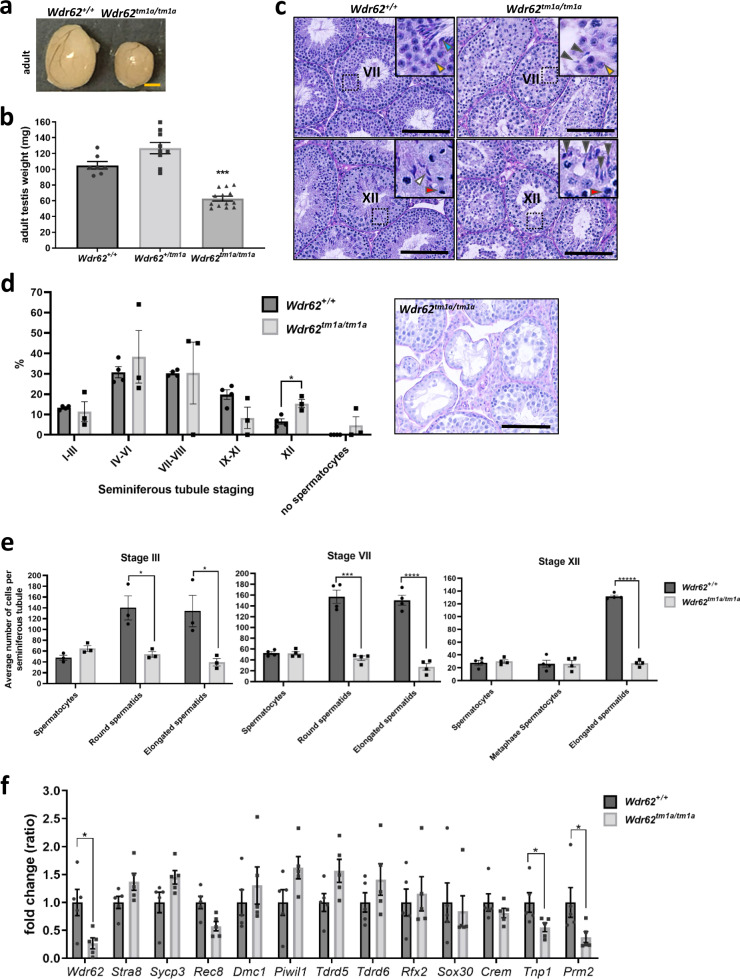
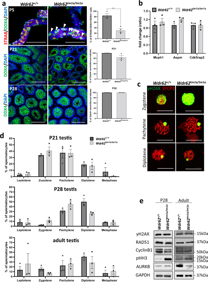
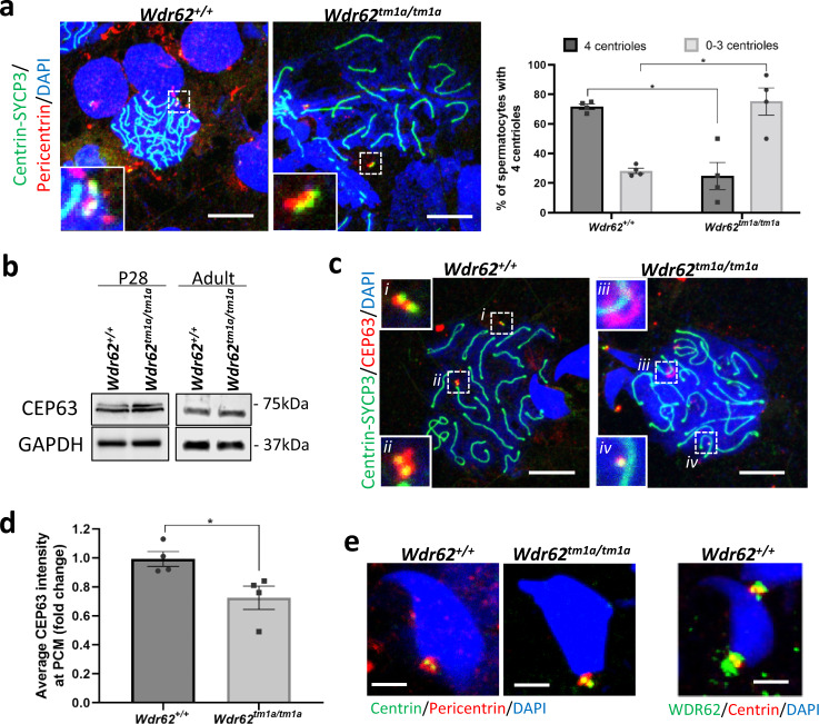
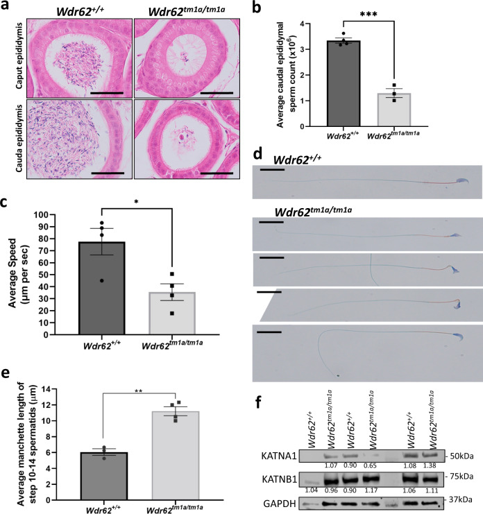
WDR62 is a scaffold protein involved in centriole duplication and spindle assembly during mitosis. Mutations in WDR62 can cause primary microcephaly and premature ovarian insufficiency. We have generated a genetrap mouse model deficient in WDR62 and characterised the developmental effects of WDR62 deficiency during meiosis in the testis. We have found that WDR62 deficiency leads to centriole underduplication in the spermatocytes due to reduced or delayed CEP63 accumulation in the pericentriolar matrix. This resulted in prolonged metaphase that led to apoptosis. Round spermatids that inherited a pair of centrioles progressed through spermiogenesis, however, manchette removal was delayed in WDR62 deficient spermatids due to delayed Katanin p80 accumulation in the manchette, thus producing misshapen spermatid heads with elongated manchettes. In mice, WDR62 deficiency resembles oligoasthenoteratospermia, a common form of subfertility in men that is characterised by low sperm counts, poor motility and abnormal morphology. Therefore, proper WDR62 function is necessary for timely spermatogenesis and spermiogenesis during male reproduction.


Conflict of interest statement

The authors declare no competing interests.

Figures

Fig. 1
Fig. 1. Wdr62tm1a/tm1a testis are smaller with reduced number of round and elongated spermatids.
a Testes from Wdr62+/+ and Wdr62tm1a/tm1a littermates collected at 7 months (adult). Scale bar represents 1 mm. b Testis weight for Wdr62+/+, Wdr62+/tm1a and Wdr62tm1a/tm1a adult testis. n = 7 Wdr62+/+, 9 Wdr62+/tm1a and 13 Wdr62tm1a/tm1a adult testes. Two-tailed unpaired Student’s t-test, ****p = 3.72 × 10−7 (see Supplementary Fig. 2c, d for P28 data). c Periodic acid Schiff-haematoxylin (PAS-H) staining shows representative stage VII and XII seminiferous tubules in Wdr62+/+ and Wdr62tm1a/tm1a adult testis. Insets show 50 μm2 magnification of selected area. Red arrowhead = dividing spermatocyte; yellow arrowhead = round spermatid; white arrowhead = elongated spermatid; blue arrowhead = spermatozoa; grey arrowhead = misshapen spermatid head. Scale bar represents 100 μm. d Quantification of PAS-H-stained adult testes sections from b. Inset: PAS-H representative image illustrating seminiferous tubules with an absence of spermatocytes, round spermatids and/or proper elongated spermatids in Wdr62tm1a/tm1a adult testis. n = 470 seminiferous tubules from three independent Wdr62+/+ adult testes; 373 tubules from three independent Wdr62tm1a/tm1a adult testes. e Quantification of the average number of spermatocytes, round spermatids and elongated spermatids per tubule in Wdr62+/+ and Wdr62tm1a/tm1a stage III, VII and XII seminiferous tubules. Mean ± SEM. Two-tailed unpaired Student’s t-test, *p = 0.020239 Stage III round spermatids, *p = 0.034665 Stage III elongated spermatids, ***p = 0.000114 Stage VII round spermatids, ****p = 0.000031 Stage VII elongated spermatids, *****p < 0.000001 Stage XII elongated spermatids. N = 15 Stage III seminiferous tubules from three adult Wdr62+/+, 14 Stage III seminiferous tubules from three adult Wdr62tm1a/tm1a, 20 Stage VII and XII seminiferous tubules from four adult Wdr62+/+, and 20 Stage VII and XII seminiferous tubules from four adult Wdr62tm1a/tm1a. f qRT-PCR of meiotic initiation (Stra8), spermatogenesis (Sycp3, Rec8, Dmc1, Piwi1, Tdrd5, Tdrd6) and round spermatid (Rfx2, Sox30, Crem, Tnp1, Prm2) markers in Wdr62+/+ and Wdr62tm1a/tm1a P28 and adult testes. N = 3 Wdr62+/+ and 5 Wdr62tm1a/tm1a P28 testes; 5 Wdr62+/+ and 5 Wdr62tm1a/tm1a adult testes. Mean ± SEM. Two-tailed unpaired Student’s t-test, *p = 0.0210 (Wdr62), p = 0.0496 (Tnp1), p = 0.0348 (Prm2) (see Supplementary Fig. 2e for P28 data).
Fig. 2
Fig. 2. Delayed meiotic initiation in Wdr62tm1a/tm1a testis.
a DDX4 (green) and STRA8 (red) whole mount immunofluorescence of P5 seminiferous tubules shows meiosis initiated (STRA8+DDX4+) germ cells in Wdr62+/+ and Wdr62tm1a/tm1a. However, some DDX4+ cells lack STRA8 nuclear staining in Wdr62tm1a/tm1a tubules (white arrowhead). Quantification of STRA8+DDX4+/total DDX4+ cells indicate reduced meiotic initiation in Wdr62tm1a/tm1a at P5. DDX4 (green) immunofluorescence of P21 and P28 testis sections show DDX4+ germ cells in seminiferous tubules in both in Wdr62+/+ and Wdr62tm1a/tm1a. Quantification of DDX4+ tubules/total seminiferous tubules in P21 and P28 testis. n = 3 Wdr62+/+ and 4 Wdr62tm1a/tm1a P5 testes, 3 Wdr62+/+ and 3 Wdr62tm1a/tm1a P21 testes, 3 Wdr62+/+ and 3 Wdr62tm1a/tm1a P28 testes. Tubules and sections were counterstained with DAPI (blue). Negative IgG controls are shown in Supplementary Fig. 7a, b. Scale bar represents 200 μm. Two-tailed unpaired Student’s t-test, **p = 0.0027. b qRT-PCR shows unaltered Mcph1, Aspm and Cdk5rap2 mRNA expression in Wdr62+/+ and Wdr62tm1a/tm1a adult testes. n = 4 Wdr62+/+ and 4 Wdr62tm1a/tm1a adult testes. c γH2AX (green) and SYCP3 (red) co-immunofluorescence on spermatocyte spreads to show different stages of meiosis prophase I. SYCP3 marks the pairing of sister chromatids, whereas γH2AX marks DNA double-strand breaks in leptotene and sex chromosomes in pachytene and diplotene. Scale bar represents 10 μm. d The number of leptotene, zygotene, pachytene, diplotene and metaphase spermatocytes from P21, P28 and adult spermatocyte spreads (based on γH2AX and SYCP3 co-staining pattern in Fig. 2c) were counted and graphed as % of total spermatocytes. n = 332 Wdr62+/+, 214 Wdr62tm1a/tm1a P21 spermatocytes; 1076 Wdr62+/+, 385 Wdr62tm1a/tm1a P28 spermatocytes; 708 Wdr62+/+, 432 Wdr62tm1a/tm1a adult spermatocytes from three independent pups per genotype per timepoint. Mean ± SEM. e Western analysis of P28 and adult testes show unchanged γH2AX, RAD51 and CyclinB1; increased pHH3 and decreased Aurora kinase B (AURKB) protein expression in adult Wdr62tm1a/tm1a testis. GAPDH was used as a loading control. See Supplementary Fig. 6 for western blots and band quantification.
Fig. 3
Fig. 3. Increased metaphase and apoptotic spermatocytes in Wdr62tm1a/tm1a testis.
a. pHH3 (green) immunofluorescence in P28 and adult testis. Negative IgG control is shown in Supplementary Fig. 7c. The percentage of pHH3-positive tubules was quantified. n = 5 Wdr62+/+ and 3 Wdr62tm1a/tm1a P28 testes; 3 Wdr62+/+ and 3 Wdr62tm1a/tm1a adult testes. Two-tailed unpaired Student’s t-test, *p = 0.0184 (P28 testis), *p = 0.0442 (adult testis). b TUNEL (green) staining in P28 and adult testis. The percentage of TUNEL-positive tubules was quantified. n = 5 Wdr62+/+ and 3 Wdr62tm1a/tm1a P28 testes; 3 Wdr62+/+ and 3 Wdr62tm1a/tm1a adult testes. Two-tailed unpaired Student’s t-test, **p = 0.0035 (P28 testis), ***p = 0.0004 (adult testis). c SYCP3 or pHH3 (red) and TUNEL (green) co-staining in the adult testis. Inset = 50 μm × 50 μm magnified area of dashed box. All sections were counterstained with DAPI (blue). All scale bars represent 100 μm.
Fig. 4
Fig. 4. Wdr62tm1a/tm1a spermatocytes exhibit centriole underduplication during meiosis.
a Centrin-SYCP3 (green) and Pericentrin (red) co-immunofluorescence on spermatocyte spreads from adult testis. Negative IgG control is shown in Supplementary Fig. 7d. The number of centrioles was quantified in pachytene and diplotene spermatocytes and graphed as % of total spermatocytes counted. In Wdr62+/+ spermatocytes, approximately 70% show four centrioles; however, this percentage is reduced to 25% in Wdr62tm1a/tm1a spermatocytes, suggesting centriole underduplication. Scale bar represents 10 μm. n = 157 spermatocytes from five independent Wdr62+/+ and 172 spermatocytes from six independent Wdr62tm1a/tm1a testis. Two-tailed Student’s t-test. *p = 0.0329. b CEP63 western blot of P28 and adult Wdr62+/+ and Wdr62tm1a/tm1a testis. GAPDH is used as loading control. See Supplementary Fig. 6 for western blots and band quantification. c Centrin-SYCP3 (green) and CEP63 (red) co-immunofluorescence on spermatocyte spreads from adult testis. Scale bar represents 10 μm. Negative IgG controls are shown in Supplementary Fig. 7e. d CEP63 intensities from c are quantified, showing approximately 20% reduction in CEP63 accumulation at the PCM in Wdr62tm1a/tm1a spermatocytes. n = 29 Wdr62+/+ and 26 Wdr62tm1a/tm1a pachytene and/or diplotene spermatocytes from three independent testis per genotype. Error bars represent SEM in a and d. Two-tailed unpaired Student’s t-test, *p = 0.0305. e [Left] Pericentrin (red) and Centrin (green) co-staining of adult Wdr62+/+ and Wdr62tm1a/tm1a spermatocyte spreads. [Right] WDR62 (green) and Centrin (Red) co-immunofluorescence of adult Wdr62+/+ spermatocyte spreads. A pair of centrioles is localised to the spermatid neck region in both Wdr62+/+ and Wdr62tm1a/tm1a samples. Cells were counterstained with DAPI (blue) in a, c, e. Scale bar represents 5 μm.
Fig. 5
Fig. 5. Wdr62tm1a/tm1a spermatozoa show oligoasthenoteratospermia.
a Haematoxylin and eosin staining of adult Wdr62+/+ and Wdr62tm1a/tm1a adult epididymis. Scare bar represents 100 μm. b Hemocytometer quantification of fixed cauda epididymal spermatozoa shows reduced sperm counts in Wdr62tm1a/tm1a. n = 4 Wdr62+/+ and 3 Wdr62tm1a/tm1a independent adult mice. Two-tailed unpaired Student’s t-test, ***p = 0.0001. c Quantification of sperm motility micrometre swam per second, which showed reduced sperm motility in Wdr62tm1a/tm1a spermatozoa. N = total 190 spermatozoa analysed from four independent Wdr62+/+ adult mice; and total 101 spermatozoa analysed from four independent Wdr62tm1a/tm1a adult mice. Two-tailed unpaired Student’s t-test, *p = 0.0180. d Sperm stain of adult cauda epididymal sperm smear, which shows normal sperm head (purple), midpiece (orange) and flagellum (green) in Wdr62+/+. However, Wdr62tm1a/tm1a spermatozoa show misshapen sperm heads, with the midpiece and flagellum appear normal. n = 3 Wdr62+/+ and 3 Wdr62tm1a/tm1a independent mice with representative images of various misshapen sperm heads shown. Scale bar represents 20 μm. e Quantification of manchette length (μm) of step 10–14 spermatids (Supplementary Fig. 5a. Acetylated α-tubulin staining of spermatids, which marks the manchette). Mean ± SEM. Two-tailed unpaired Student’s t-test, **p = 0.0010. n = 11 Wdr62+/+ and 49 Wdr62tm1a/tm1a step 10–14 spermatids from four independent adult mice per genotype. f Western analysis shows unaltered Katanin p60 (KATNA1) and Katanin p80 (KATNB1) protein expression in Wdr62+/+ and Wdr62tm1a/tm1a adult testis. Normalised band quantification as shown. GAPDH was used as a loading control.
Fig. 6
Fig. 6. Wdr62tm1a/tm1a spermatids exhibit defective manchette removal.
a Katanin p60 (KATNA1, green) and acetylated α-tubulin (red) co-immunofluorescence on adult spermatocyte spreads. Representative images of co-staining in step 8–14 spermatids are shown. Scale bar represents 5 μm. b Katanin p80 (KATNB1, green) and acetylated α-tubulin (red) co-immunofluorescence on adult spermatocyte spreads. Representative images of staining in step 8–14 spermatids are shown. All spermatids are counterstained with DAPI (blue) in (a) and (b). Scale bars represent 5 μm. Negative IgG controls for (a) and (b) are shown in Supplementary Fig. 7f.

References

    1. Cobb J, Handel MA. Dynamics of meiotic prophase I during spermatogenesis: from pairing to division. Semin. Cell Dev. Biol. 1998;9:445–450. doi: 10.1006/scdb.1998.0202. - DOI - PubMed
    1. Breslow DK, Holland AJ. Mechanism and regulation of centriole and cilium biogenesis. Annu. Rev. Biochem. 2019;88:691–724. doi: 10.1146/annurev-biochem-013118-111153. - DOI - PMC - PubMed
    1. Peters N, et al. Control of mitotic and meiotic centriole duplication by the Plk4-related kinase ZYG-1. J. Cell Sci. 2010;123:795–805. doi: 10.1242/jcs.050682. - DOI - PMC - PubMed
    1. Farag HG, et al. Abnormal centrosome and spindle morphology in a patient with autosomal recessive primary microcephaly type 2 due to compound heterozygous WDR62 gene mutation. Orphanet J. Rare Dis. 2013;8:178. doi: 10.1186/1750-1172-8-178. - DOI - PMC - PubMed
    1. Yu TW, et al. Mutations in WDR62, encoding a centrosome-associated protein, cause microcephaly with simplified gyri and abnormal cortical architecture. Nat. Genet. 2010;42:1015–1020. doi: 10.1038/ng.683. - DOI - PMC - PubMed

Publication types

LinkOut - more resources