

Clinical and radiographic evaluation of triple antibiotic paste pulp therapy compared to Vitapex pulpectomy in non-vital primary molars
- PMID: 34060250
- PMCID: PMC8543458
- DOI: 10.1002/cre2.434
Clinical and radiographic evaluation of triple antibiotic paste pulp therapy compared to Vitapex pulpectomy in non-vital primary molars
Abstract
Objectives: This study compared and evaluated the clinical and radiographic efficacy of non-instrumentation triple antibiotic paste pulp therapy and Vitapex pulpectomy in non-vital primary molars.
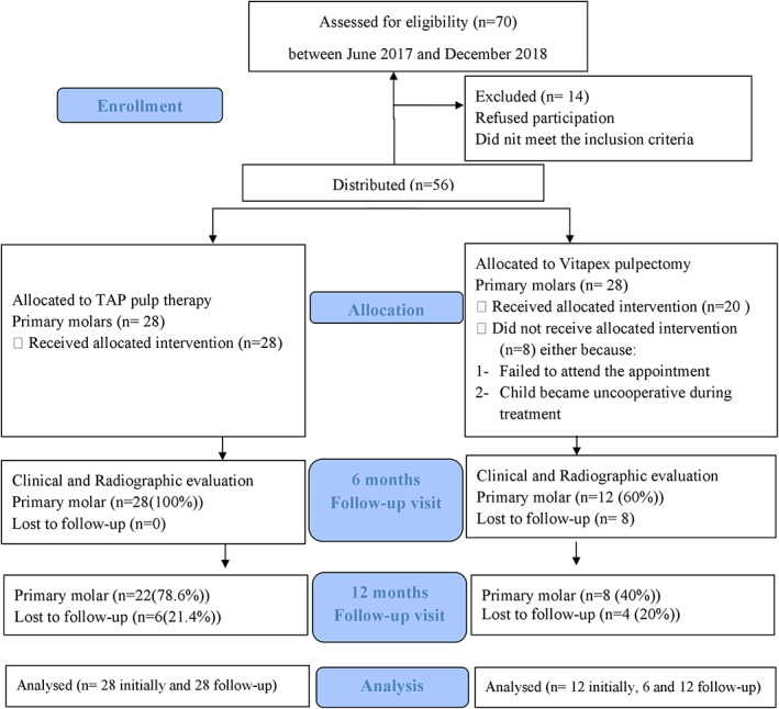
Material and methods: Healthy, 5-9 years old children with at least one non-vital primary molar were included in the study. Molars were divided into two groups based on the subject's cooperation level. In the first group, molars received triple antibiotic paste, and a second group received Vitapex pulpectomy followed by a stainless-steel crown. Triple antibiotic paste was freshly prepared and proportioned in equal parts by volume (metronidazole, minocycline, and ciprofloxacin = 1:1:1) before the scheduled treatment. A clinical and radiographic examination was performed by two trained and calibrated pediatric dentists at the pre-operative baseline and the 6- and 12-month follow-up visits.
Results: A total of 28 molars received triple antibiotic paste pulp therapy and 20 received Vitapex pulpectomy. At the 6-month follow-up, the success rate among the molars in the triple antibiotic paste group was clinically (92.85%) and radiographically (85.71%) higher compared to the Vitapex group (91.67%, 62.50% respectively) with p = 0.89 and 0.55 respectively. At the 12-month follow-up, the molars in the triple antibiotic paste group showed lower clinical (95.45%) but higher radiographic success rate (72.73%) compared to the Vitapex group (100% and 62.50%) with (p = 0.85 and 0.47) respectively. None of the differences were statistically significant.
Conclusions: Both triple antibiotic paste and Vitapex can be clinically and radiographically effective in treating non-vital primary molars.
Keywords: deciduous teeth; endodontic treatment; primary molars and pulpectomy..
© 2021 The Authors. Clinical and Experimental Dental Research published by John Wiley & Sons Ltd.
Conflict of interest statement
The authors declare no potential conflict of interest.
Figures


References
-
- Ahmed, H. (2013). Anatomical challenges, electronic working length determination and current developments in root canal preparation of primary molar teeth. International Endodontic Journal, 46(11), 1011–1022. - PubMed
-
- Ahmed, H. M. A. (2014). Pulpectomy procedures in primary molar teeth. European Journal of General Dentistry, 3(1), 3.
-
- American Academy of Pediatric Dentistry . (2017). Prescribing dental radiographs for infants, children, adolescents, and individuals with special health care needs. Pediatric Dentistry, 39(6), 205–207. - PubMed
-
- American Academy of Pediatric Dentistry Clinical Affairs Committee–Developing Dentition Subcommittee; American Academy of Pediatric Dentistry Council on Clinical Affairs . (2005). Guideline on management of the developing dentition and occlusion in pediatric dentistry. Pediatric Dentistry, 27(7 Suppl), 143. - PubMed
-
- Asl Aminabadi, N. , Satrab, S. , Najafpour, E. , Samiei, M. , Jamali, Z. , & Shirazi, S. (2016). A randomized trial of direct pulp capping in primary molars using MTA compared to 3Mixtatin: A novel pulp capping biomaterial. International Journal of Paediatric Dentistry, 26(4), 281–290. 10.1111/ipd.12196 - DOI - PubMed
MeSH terms
Substances
LinkOut - more resources
Full Text Sources
Medical
