

Convalescent-Phase Sera and Vaccine-Elicited Antibodies Largely Maintain Neutralizing Titer against Global SARS-CoV-2 Variant Spikes
- PMID: 34060334
- PMCID: PMC8262901
- DOI: 10.1128/mBio.00696-21
Convalescent-Phase Sera and Vaccine-Elicited Antibodies Largely Maintain Neutralizing Titer against Global SARS-CoV-2 Variant Spikes
Abstract
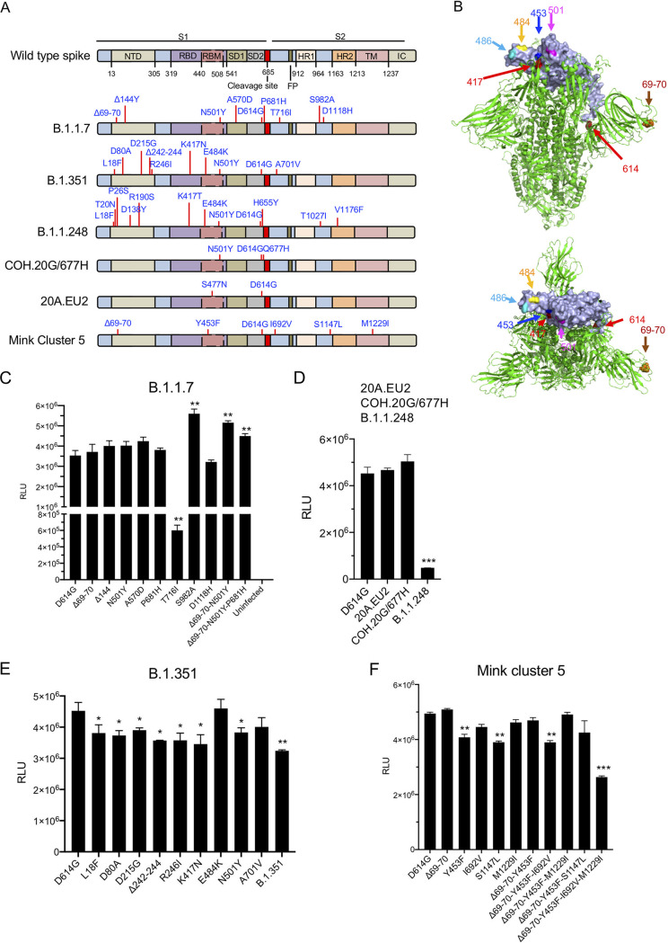
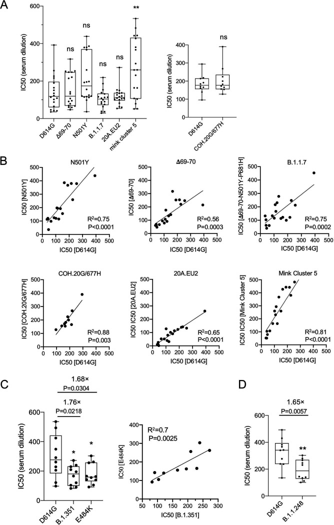
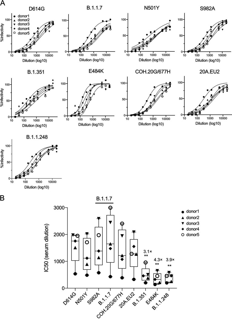
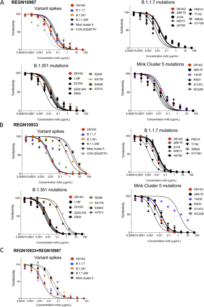
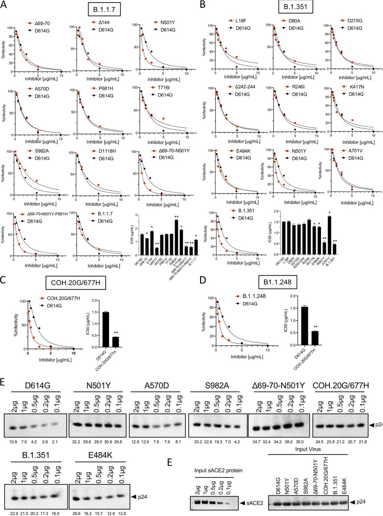
The increasing prevalence of severe acute respiratory syndrome coronavirus 2 (SARS-CoV-2) variants with spike protein mutations raises concerns that antibodies elicited by natural infection or vaccination and therapeutic monoclonal antibodies will become less effective. We show that convalescent-phase sera neutralize pseudotyped viruses with the B.1.1.7, B.1.351, B.1.1.248, COH.20G/677H, 20A.EU2, and mink cluster 5 spike proteins with only a minor loss in titer. Similarly, antibodies elicited by Pfizer BNT162b2 vaccination neutralized B.1.351 and B.1.1.248 with only a 3-fold decrease in titer, an effect attributable to E484K. Analysis of the Regeneron monoclonal antibodies REGN10933 and REGN10987 showed that REGN10933 has lost neutralizing activity against the B.1.351 and B.1.1.248 pseudotyped viruses, and the cocktail is 9- to 15-fold decreased in titer. These findings suggest that antibodies elicited by natural infection and by the Pfizer vaccine will maintain protection against the B.1.1.7, B.1.351, and B.1.1.248 variants but that monoclonal antibody therapy may be less effective for patients infected with B.1.351 or B.1.1.248 SARS-CoV-2. IMPORTANCE The rapid evolution of SARS-CoV-2 variants has raised concerns with regard to their potential to escape from vaccine-elicited antibodies and anti-spike protein monoclonal antibodies. We report here on an analysis of sera from recovered patients and vaccinated individuals and on neutralization by Regeneron therapeutic monoclonal antibodies. Overall, the variants were neutralized nearly as well as the wild-type pseudotyped virus. The B.1.351 variant was somewhat resistant to vaccine-elicited antibodies but was still readily neutralized. One of the two Regeneron therapeutic monoclonal antibodies seems to have lost most of its activity against the B.1.351 variant, raising concerns that the combination therapy might be less effective for some patients. The findings should alleviate concerns that vaccines will become ineffective but suggest the importance of continued surveillance for potential new variants.
Keywords: 20A.EU2; B.1.1.248; B.1.1.7; B.1.351; COH.20G/677H; Pfizer BNT162b2; REGN10933; REGN10987; SARS-CoV-2; mink cluster 5; neutralization; spike protein.
Figures
Update of
-
Neutralization of viruses with European, South African, and United States SARS-CoV-2 variant spike proteins by convalescent sera and BNT162b2 mRNA vaccine-elicited antibodies.bioRxiv [Preprint]. 2021 Feb 7:2021.02.05.430003. doi: 10.1101/2021.02.05.430003. bioRxiv. 2021. Update in: mBio. 2021 Jun 29;12(3):e0069621. doi: 10.1128/mBio.00696-21. PMID: 33564768 Free PMC article. Updated. Preprint.
Similar articles
-
B.1.526 SARS-CoV-2 Variants Identified in New York City are Neutralized by Vaccine-Elicited and Therapeutic Monoclonal Antibodies.mBio. 2021 Aug 31;12(4):e0138621. doi: 10.1128/mBio.01386-21. Epub 2021 Jul 27. mBio. 2021. PMID: 34311587 Free PMC article.
-
Neutralization of viruses with European, South African, and United States SARS-CoV-2 variant spike proteins by convalescent sera and BNT162b2 mRNA vaccine-elicited antibodies.bioRxiv [Preprint]. 2021 Feb 7:2021.02.05.430003. doi: 10.1101/2021.02.05.430003. bioRxiv. 2021. Update in: mBio. 2021 Jun 29;12(3):e0069621. doi: 10.1128/mBio.00696-21. PMID: 33564768 Free PMC article. Updated. Preprint.
-
Escape from neutralizing antibodies by SARS-CoV-2 spike protein variants.Elife. 2020 Oct 28;9:e61312. doi: 10.7554/eLife.61312. Elife. 2020. PMID: 33112236 Free PMC article.
-
Variants of SARS-CoV-2, their effects on infection, transmission and neutralization by vaccine-induced antibodies.Eur Rev Med Pharmacol Sci. 2021 Sep;25(18):5857-5864. doi: 10.26355/eurrev_202109_26805. Eur Rev Med Pharmacol Sci. 2021. PMID: 34604978 Review.
-
The emergence of SARS-CoV-2 variants threatens to decrease the efficacy of neutralizing antibodies and vaccines.Biochem Soc Trans. 2021 Dec 17;49(6):2879-2890. doi: 10.1042/BST20210859. Biochem Soc Trans. 2021. PMID: 34854887 Free PMC article. Review.
Cited by
-
Comparative analysis of human immune responses following SARS-CoV-2 vaccination with BNT162b2, mRNA-1273, or Ad26.COV2.S.NPJ Vaccines. 2022 Jul 6;7(1):77. doi: 10.1038/s41541-022-00504-x. NPJ Vaccines. 2022. PMID: 35794181 Free PMC article.
-
A spike-targeting bispecific T cell engager strategy provides dual layer protection against SARS-CoV-2 infection in vivo.Commun Biol. 2023 Jun 1;6(1):592. doi: 10.1038/s42003-023-04955-3. Commun Biol. 2023. PMID: 37264086 Free PMC article.
-
Current Updates on Variants of SARS-CoV- 2: Systematic Review.Health Sci Rep. 2024 Nov 4;7(11):e70166. doi: 10.1002/hsr2.70166. eCollection 2024 Nov. Health Sci Rep. 2024. PMID: 39502131 Free PMC article. Review.
-
Single-virus tracking reveals variant SARS-CoV-2 spike proteins induce ACE2-independent membrane interactions.Sci Adv. 2022 Dec 9;8(49):eabo3977. doi: 10.1126/sciadv.abo3977. Epub 2022 Dec 9. Sci Adv. 2022. PMID: 36490345 Free PMC article.
-
Neutralization assays for SARS-CoV-2: Implications for assessment of protective efficacy of COVID-19 vaccines.Indian J Med Res. 2022 Jan;155(1):105-122. doi: 10.4103/ijmr.ijmr_2544_21. Indian J Med Res. 2022. PMID: 35859437 Free PMC article. Review.
References
-
- Isabel S, Graña-Miraglia L, Gutierrez JM, Bundalovic-Torma C, Groves HE, Isabel MR, Eshaghi A, Patel SN, Gubbay JB, Poutanen T, Guttman DS, Poutanen SM. 2020. Evolutionary and structural analyses of SARS-CoV-2 D614G spike protein mutation now documented worldwide. Sci Rep 10:14031. doi:10.1038/s41598-020-70827-z. - DOI - PMC - PubMed
-
- Zhang L, Jackson CB, Mou H, Ojha A, Peng H, Quinlan BD, Rangarajan ES, Pan A, Vanderheiden A, Suthar MS, Li W, Izard T, Rader C, Farzan M, Choe H. 2020. SARS-CoV-2 spike-protein D614G mutation increases virion spike density and infectivity. Nat Commun 11:6013. doi:10.1038/s41467-020-19808-4. - DOI - PMC - PubMed
-
- Volz E, Mishra S, Chand M, Barrett JC, Johnson R, Geidelberg L, Hinsley WR, Laydon DJ, Dabrera G, O’Toole Á, Amato R, Ragonnet-Cronin M, Harrison I, Jackson B, Ariani CV, Boyd O, Loman L, McCrone JT, Gonçalves S, Jorgensen D, Myers R, Hill V, Jackson DK, Gaythorpe K, Groves N, Sillitoe J, Kwiatkowski DP, COG-UK, Flaxman S, Ratmann O, Bhatt S, Hopkins S, Gandy A, Rambaut A, Ferguson NM. 2021. Transmission of SARS-CoV-2 lineage B.1.1.7 in England: insights from linking epidemiological and genetic data. medRxiv 10.1101/2020.12.30.20249034. - DOI
Publication types
MeSH terms
Substances
Grants and funding
LinkOut - more resources
Full Text Sources
Medical
Molecular Biology Databases
Research Materials
Miscellaneous
