. 2021 Jun 1;131(11):e141810.
doi: 10.1172/JCI141810.

Lung-resident memory B cells protect against bacterial pneumonia

Affiliations

Lung-resident memory B cells protect against bacterial pneumonia

Kimberly A Barker et al. J Clin Invest. .

Abstract

Lung-resident memory B cells (BRM cells) are elicited after influenza infections of mice, but connections to other pathogens and hosts - as well as their functional significance - have yet to be determined. We postulate that BRM cells are core components of lung immunity. To test this, we examined whether lung BRM cells are elicited by the respiratory pathogen pneumococcus, are present in humans, and are important in pneumonia defense. Lungs of mice that had recovered from pneumococcal infections did not contain organized tertiary lymphoid organs, but did have plasma cells and noncirculating memory B cells. The latter expressed distinctive surface markers (including CD69, PD-L2, CD80, and CD73) and were poised to secrete antibodies upon stimulation. Human lungs also contained B cells with a resident memory phenotype. In mice recovered from pneumococcal pneumonia, depletion of PD-L2+ B cells, including lung BRM cells, diminished bacterial clearance and the level of pneumococcus-reactive antibodies in the lung. These data define lung BRM cells as a common feature of pathogen-experienced lungs and provide direct evidence of a role for these cells in pulmonary antibacterial immunity.

Keywords: Adaptive immunity; Bacterial infections; Immunology; Pulmonology.


Conflict of interest statement

Conflict of interest: The authors have declared that no conflict of interest exists.

Figures

Figure 1
Figure 1. Pneumococcal exposures provide lung protection without extensive changes to the blood transcriptome.
(A) B6 mice were exposed to i.t. or i.n. pneumococcus (Sp19F) or saline in the left lung lobe twice with a 1-week interval, then allowed to recover for at least 4 weeks, at which point they were referred to as “experienced” or “naive,” respectively. (B) Experienced and naive mice were challenged with i.t. pneumococcus (Sp3) for 24 hours before lung bacterial burdens were assessed (Mann-Whitney U test, *P = 0.0006). Principal component analysis (C) and read counts (D) from RNA-Seq of whole blood collected from the same naive and experienced mice as in B prior to Sp3 challenge. Data point size in C is proportional to the 24-hour lung Sp3 CFU of each mouse in B. Each point in D represents 1 gene.
Figure 2
Figure 2. Pneumococcal exposure elicits clusters of EV memory B cells in the lung.
Immunofluorescence staining (20×) for B220 (red), CD4 (green), and nuclei (DAPI, blue) of representative naive (A) and experienced (B) lungs. H&E staining of representative naive (C) and experienced (D) lungs. (E and F) Left panels: Magnified view of 2 immune cell clusters in D. Right panels: Immunohistochemical staining for CD19 (red) and CD4 (cyan) in serial sections of the same lungs imaged in the left panels. Scale bars: 75 μm (A and B); 500 μm (C and D); 100 μm (E and F). (G) Representative gating of i.v. CD45+CD19+ cells (IV B cells) and i.v. CD45CD19+ cells (EV B cells) in lungs of naive and experienced mice. (H) IgD expression on IV and EV B cells in experienced lungs (left) and percentages of IV and EV B cells that are IgD in experienced lungs (right; Mann-Whitney U test, *P < 0.0001). (I) Percentages of IgD+ and IgD B cells among live lung cells in the IV and EV compartments of naive and experienced mice analyzed between 4 and 12 weeks after the previous Sp19F exposure (2-way ANOVA, *P = 0.029, **P = 0.0006, ***P < 0.0001). (J) IgD EV B cells in naive and experienced lungs at the indicated times after lung exposure (2-way ANOVA, *P = 0.01, **P = 0.0054, ***P < 0.0001). (K) CD38 expression on EV IgD B cells (each red curve from 1 of 5 different mice) and on EV CD45 cells (shaded curve, representative). A, airway; pa, pulmonary artery.
Figure 3
Figure 3. Pneumococcus-specific plasma cells are found in both bone marrow and lungs of experienced mice.
(A) Plasma of naive and experienced mice was assessed for Sp3-reactive antibodies via ELISA. n = 3 for naive mice and n = 6 or more for experienced mice. (B) Pneumococcus-specific antibodies in supernatants from overnight cultures of naive or experienced bone marrow cells. n = 4 per group. 2-way ANOVA, **P = 0.0079, ***P < 0.0001. Total IgG and IgA ELISpot images (C) and counts (D) of EV lung plasma cells (PC) (Mann-Whitney U tests, **P = 0.0037, ***P = 0.0004). Vertical line in C separates nonconsecutive wells from the same plate. Representative flow plots (E) and quantification (F) of EV plasma cells (CD138+CD38lo) and CD38+CD138 cells from an experienced mouse lung (Mann-Whitney U test, *P = 0.032). (G) Representative intracellular Blimp-1 expression in EV CD38loCD138+ and CD38+CD138 lung cells. (H) BALF of naive and experienced mice was assessed for Sp3-reactive antibodies via ELISA. n = 4 for naive mice and n = 7 or more for experienced mice.
Figure 4
Figure 4. Lung B cells in experienced mice are resident.
(A) Representative flow cytometry plots showing expression of CD69, CD11a, CD62L, and CD44 on IV and EV IgD lung B cells in experienced mice. (B) Percentage of CD69+ of EV IgD B cells in the exposed left lung lobe and control right lung lobes of naive and experienced mice (1-way ANOVA, *P = 0.0054, **P = 0.0021, ***P < 0.0001). (C) Median fluorescence intensity (MFI) of CD11a on EV IgD B cells in the exposed left lung lobe and control right lung lobes of naive and experienced mice (1-way ANOVA, *P = 0.0047, **P = 0.0017, ***P < 0.0001). (D) Fold change in EV IgD lung B cells between saline-treated and experienced mice in the exposed left lung lobe and unexposed control right lobe. Each dot represents the average fold change in 1 experiment including at least 3 naive and 3 experienced mice (Mann-Whitney U test, *P = 0.036). (E) Binding of a fluorescent anti-CD20 antibody to lung B cells from mice treated 2 weeks previously with isotype control IgG or anti-CD20 (5D2 clone). FMO, fluorescence minus one. Representative flow cytometry plots (F) and quantification CD19+ cells (G) in lung compartments 4 days or 2 weeks after anti-CD20 or IgG treatment of experienced mice (Kruskal-Wallis test, *P = 0.002).
Figure 5
Figure 5. Human lungs are enriched for B cells bearing a resident memory phenotype.
(A) Normal tissue from wedge resection or biopsy samples from humans with lung cancer were collagenase digested and analyzed via flow cytometry using the gating scheme shown. (BF) Various cell surface phenotypes of B and T cells in the human lung digests. (B) Percentage of CD4+ and CD19+ cells among live, single cells (Mann-Whitney U test, *P = 0.0023). (C) Percentage of CD27+ cells among all CD19+ cells and among naive (CD19+IgD+) B cells (Mann-Whitney U test, *P = 0.0025). (D) Percentage of CD69+ cells among CD4+ cells, memory B cells (CD19+CD27+), and naive B cells (Kruskal-Wallis test, *P = 0.021, **P = 0.049). (E) Percentage of memory B cells that are class switched (Class sw.) and percentage that are IgM+ (Mann-Whitney U test, *P = 0.0079). (F) Percentage of resident memory B cells (CD27+CD69+CD19+) negative for the B cell activation marker CD83 and for CD38.
Figure 6
Figure 6. Lung BRM cells are poised to secrete antibody.
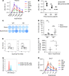
(A) Distribution of naive, IgM+IgD, and class-switched B cells in experienced lungs and spleens (2-way ANOVA comparing isotypes across compartments, n = 3 for spleen, 5 for lung; *P = 0.0022 vs. spleen, P = 0.0009 vs. IV lung; **P < 0.0001 vs. either compartment). (B) Experienced EV lung IgMIgD B cell isotypes. n = 5. Representative flow plots from experienced lungs showing MBC marker expression on IgD EV B cells (C) and coexpression (D) as quantified in E, where numbers of triple-negative (TN), single-positive (SP), and double- or triple-positive (DP/TP) B cells are shown (2-way ANOVA comparing each marker category between naive and experienced mice, *P < 0.0001). Total IgG ELISpot image (F) and counts (G) of spleen and EV lung B cells after ex vivo culture (Kruskal-Wallis test for each organ, *P = 0.028, **P = 0.0035 for spleen, P = 0.006 for lung). Vertical lines in F separate images within a group obtained from different plates. (H) Pneumococcus-specific antibody levels in ex vivo culture supernatants from F and G (Kruskal-Wallis test for each isotype within each organ, *P = 0.0039 for IgG, P = 0.0064 for IgM). (I) Pneumococcus-specific antibody in experienced BALF at baseline or after Sp3 infection (Mann-Whitney U test for each isotype, n = 15 for baseline, n = 8 for 96 hours; *P = 0.0018, **P = 0.0007, ***P < 0.0001).
Figure 7
Figure 7. Systemic B cell immunity may contribute to, but is not required for, serotype-independent lung antipneumococcal immunity in experienced mice.
(A) Twenty-four-hour lung Sp3 burdens in naive and experienced B6 or μMT mice (2-way ANOVA, *P = 0.013, **P = 0.0004, ***P < 0.0001). (B) Weight loss after Sp3 challenge in experienced B6 and μMT mice (2-way ANOVA comparing mouse strains within each time point, n = 16 B6 and n = 9 μMT; *P = 0.0004). (C) Plasma of experienced or naive mice was used to pre-opsonize Sp3 prior to bacterial instillation in naive mice and determination of 24-hour lung CFU (Mann-Whitney U test, *P = 0.01). Twenty-four-hour lung Sp3 CFU (D; no significant differences by 2-way ANOVA) and plasma antipneumococcal antibody titers (E) were determined in experienced mice that were splenectomized or given sham surgery 3 weeks prior to Sp3 infection (for E, Mann-Whitney U test for each isotype, *P = 0.046). (F) Twenty-four-hour lung Sp3 burdens in naive and experienced mice treated 4 days prior with isotype control or anti-CD20 (2-way ANOVA, *P = 0.0024 for IgG, P = 0.0027 for anti-CD20). LoD, limit of detection.
Figure 8
Figure 8. Lung PD-L2+ MBC are required for optimal serotype-independent antipneumococcal lung immunity and local IgG production.
(A) Timeline of treatments in PZTD mice. Representative plots of EV lung B cells in naive and experienced Cre+ mice (B) and in experienced Cre+ mice with or without DT (C). (D) Quantification of PD-L2+ EV B cells in lungs of experienced Cre and Cre+ mice with or without DT (2-way ANOVA, n = 7 for vehicle-treated Cre, n = 3 for DT-treated Cre, n = 8 for vehicle-treated Cre+, and 10 for DT-treated Cre+ mice; *P = 0.029). (E) Weight loss after Sp3 challenge in DT-treated experienced Cre and Cre+ mice (2-way ANOVA comparing genotypes at each time point, n = 11 per genotype; *P = 0.0093). (F) Ninety-six-hour lung Sp3 burdens in DT-treated experienced Cre and Cre+ mice (Mann-Whitney U test, *P = 0.0004). Squares, female; circles, male for Cre+ group. One male mouse (identified with an X) died shortly before lung harvest. IL-17 (G) and IFN-γ (H) levels in lung homogenates of DT-treated experienced PZTD mice after 24 or 96 hours of Sp3 infection (no significant differences by Mann-Whitney U tests within each time point). (I) Plasma of mice from F was assessed for Sp3-reactive antibodies via ELISA (no significant differences by Mann-Whitney U test within each isotype). (J) BALF from experienced DT-treated Cre and Cre+ PZTD mice was collected in a single experiment 96 hours after i.n. Sp3 infection and assessed for Sp3-reactive antibodies via ELISA. Two-way ANOVAs, n = 6 for Cre mice and n = 4 for Cre+ mice; *P = 0.048. hpi, hours after infection.

References

    1. Beura LK, et al. Normalizing the environment recapitulates adult human immune traits in laboratory mice. Nature. 2016;532(7600):512–516. doi: 10.1038/nature17655. - DOI - PMC - PubMed
    1. Sathaliyawala T, et al. Distribution and compartmentalization of human circulating and tissue-resident memory T cell subsets. Immunity. 2013;38(1):187–197. doi: 10.1016/j.immuni.2012.09.020. - DOI - PMC - PubMed
    1. Masopust D, Soerens AG. Tissue-resident T cells and other resident leukocytes. Annu Rev Immunol. 2019;37:521–546. doi: 10.1146/annurev-immunol-042617-053214. - DOI - PMC - PubMed
    1. Turner DL, et al. Lung niches for the generation and maintenance of tissue-resident memory T cells. Mucosal Immunol. 2014;7(3):501–510. doi: 10.1038/mi.2013.67. - DOI - PMC - PubMed
    1. Reboldi A, Cyster JG. Peyer’s patches: organizing B-cell responses at the intestinal frontier. Immunol Rev. 2016;271(1):230–245. doi: 10.1111/imr.12400. - DOI - PMC - PubMed

Publication types

Substances