Review
. 2021 Jun 1;131(11):e148671.
doi: 10.1172/JCI148671.

Ironing out mechanisms of iron homeostasis and disorders of iron deficiency

Review

Ironing out mechanisms of iron homeostasis and disorders of iron deficiency

Navid Koleini et al. J Clin Invest. .

Abstract

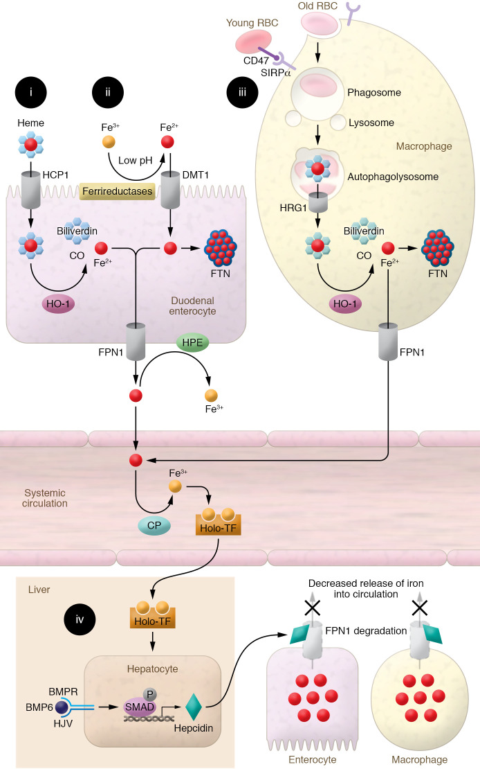
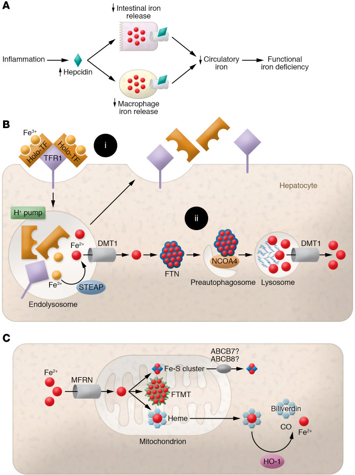
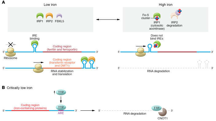
Iron plays an important role in mammalian physiological processes. It is a critical component for the function of many proteins, including enzymes that require heme and iron-sulfur clusters. However, excess iron is also detrimental because of its ability to catalyze the formation of reactive oxygen species. As a result, cellular and systemic iron levels are tightly regulated to prevent oxidative damage. Iron deficiency can lead to a number of pathological conditions, the most prominent being anemia. Iron deficiency should be corrected to improve adult patients' symptoms and to facilitate normal growth during fetal development and childhood. However, inappropriate use of intravenous iron in chronic conditions, such as cancer and heart failure, in the absence of clear iron deficiency can lead to unwanted side effects. Thus, this form of therapy should be reserved for certain patients who cannot tolerate oral iron and need rapid iron replenishment. Here, we will review cellular and systemic iron homeostasis and will discuss complications of iron deficiency.


Conflict of interest statement

Conflict of interest: Hossein Ardehali receives income as an expert witness.

Figures

Figure 1
Figure 1. Depiction of the mechanism of systemic iron regulation.
(i) Heme-bound iron is absorbed into duodenal enterocytes, possibly via HCP1. Enterocyte HO-1 releases iron from heme’s porphyrin ring, producing Fe2+, biliverdin, and carbon monoxide. Fe2+ iron is then exported into the circulation by FPN1. (ii) Absorbed Fe3+ is reduced to Fe2+ at the brush border by low pH and ferrireductases, enabling its transport by DMT1 into duodenal enterocytes. Fe2+ is either bound to FTN within the enterocyte, limiting the intracellular pool of free iron, or released into the circulation by FPN1, where it is oxidized to Fe3+ by hephaestin (HPE) and ceruloplasmin (CP) and bound to TF for transport. (iii) Macrophages identify senescent RBCs no longer expressing CD47 via SIRPα and recycle RBC iron through phagocytosis. Upon fusion of phagosomes with lysosomes, heme is released from hemoglobin and transported to the cytosol via HRG1. In the cytosol, heme-bound iron is extracted by HO-1 and exported by FPN1. Fe2+ is then oxidized to Fe3+ by CP and binds to TF for transport. (iv) When systemic iron levels are sufficiently replete, hepcidin is produced by the liver, binds to FPN1 on macrophages and enterocytes, and promotes its degradation. This prevents intestinal and macrophage iron release into the circulation. Binding of BMP6 to BMP receptor and its coreceptor HJV activates SMAD signaling and promotes transcription of hepcidin.
Figure 2
Figure 2. Depiction of the mechanism of ACD and cellular and mitochondrial iron regulation.
(A) Graphic of pathways leading to functional iron deficiency (ID). Inflammation increases hepcidin expression, inhibiting intestinal and macrophage iron release into the circulation. This simultaneously decreases systemic iron bioavailability and traps iron within tissues, leading to functional ID despite normal iron storage. (B) (i) Iron bound to TF is absorbed by hepatocytes via binding to TFR1 and subsequent receptor-mediated endocytosis. Iron is reduced from Fe3+ to Fe2+ and released from TFR1-TF in acidified endolysosomes by the action of the STEAP family of ferrireductases. Fe2+ is then exported to the cytosol via DMT1, while TFR1 and TF are recycled back to the cell surface. (ii) Within the cytosol, iron is stored by binding to FTN to reduce free-iron toxicity. Iron is released from FTN via receptor (NCOA4)-mediated autophagy and is exported to the cytosol via lysosomal DMT1. (C) Elemental iron is imported across the mitochondrial membrane by MFRN1/2. Synthesis of Fe-S clusters and heme occurs primarily within mitochondria. Fe-S clusters are exported into the cytosol via unknown mechanisms, but may require ABCB7 and ABCB8.
Figure 3
Figure 3. Regulation of cellular iron by the IRP system and TTP.
(A) IRPs dynamically bind IREs in the UTRs of target mRNAs to regulate the expression of proteins important in iron metabolism. When iron is sufficient, IRP1 and IRP2 do not bind mRNA because (a) IRP1 binds Fe-S clusters, preventing IRP1 from binding to the IREs on target mRNAs, and (b) IRP2 is degraded in a process dependent on FBXL5. In ID, binding of IRPs to IREs located at the 5′-UTR of the transcripts causes steric blockage of ribosomal entry and prevention of translation (FPN, FTN). Binding of IRPs to IREs located on the 3′-UTR of target mRNAs increases stability of the transcript and thus increases translation (TFR1, DMT1). Accordingly, this leads to upregulated translation of iron acquisition proteins such as TFR1 and DMT1 and downregulation of proteins that bind or export iron such as FTN or FPN. (B) In severe ID, iron is preserved for vital processes via a mechanism called the iron conservation pathway. Critically low iron induces upregulation of TTP that binds AU-rich elements (AREs) in the 3′-UTR of target mRNAs. Under critically low iron conditions, ARE-bound TTP recruits CNOT1, a member of the CCR4-NOT deadenylase complex, to promote degradation of target mRNAs. Multiple TTP target mRNAs encode iron-binding proteins that could sponge the limited iron available in the cell, such as mitochondrial Fe-S–containing proteins, necessitating their translational downregulation in severe ID.

Comment in

References

    1. Wallace DF. The regulation of iron absorption and homeostasis. Clin Biochem Rev. 2016;37(2):51–62. - PMC - PubMed
    1. Meng Y, et al. Jumonji domain-containing protein family: the functions beyond lysine demethylation. J Mol Cell Biol. 2018;10(4):371–373. doi: 10.1093/jmcb/mjy010. - DOI - PMC - PubMed
    1. Shen J, et al. Structure and mechanism of a unique diiron center in mammalian Stearoyl-CoA desaturase. J Mol Biol. 2020;432(18):5152–5161. doi: 10.1016/j.jmb.2020.05.017. - DOI - PMC - PubMed
    1. Braymer JJ, Lill R. Iron-sulfur cluster biogenesis and trafficking in mitochondria. J Biol Chem. 2017;292(31):12754–12763. doi: 10.1074/jbc.R117.787101. - DOI - PMC - PubMed
    1. Ganz T. Systemic iron homeostasis. Physiol Rev. 2013;93(4):1721–1741. doi: 10.1152/physrev.00008.2013. - DOI - PubMed

Publication types