Review
. 2021 Apr;135(2):165-173.
doi: 10.1037/bne0000442.

Defining an orbitofrontal compass: Functional and anatomical heterogeneity across anterior-posterior and medial-lateral axes

Affiliations
Review

Defining an orbitofrontal compass: Functional and anatomical heterogeneity across anterior-posterior and medial-lateral axes

Ines V Barreiros et al. Behav Neurosci. 2021 Apr.

Abstract

The orbitofrontal cortex (OFC) plays a critical role in the flexible control of behaviors and has been the focus of increasing research interest. However, there have been a number of controversies around the exact theoretical role of the OFC. One potential source of these issues is the comparison of evidence from different studies, particularly across species, which focus on different specific sub-regions within the OFC. Furthermore, there is emerging evidence that there may be functional diversity across the OFC which may account for these theoretical differences. Therefore, in this review we consider evidence supporting functional heterogeneity within the OFC and how it relates to underlying anatomical heterogeneity. We highlight the importance of anatomical and functional distinctions within the traditionally defined OFC subregions across the medial-lateral axis, which are often not differentiated for practical and historical reasons. We then consider emerging evidence of even finer-grained distinctions within these defined subregions along the anterior-posterior axis. These fine-grained anatomical considerations reveal a pattern of dissociable, but often complementary functions within the OFC. (PsycInfo Database Record (c) 2021 APA, all rights reserved).


Figures
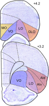
Figure 1
Figure 1. Classical Parcellation of Rat OFC
Note. Currently recognized OFC subdivisions include MO, VO, LO, DLO, and AI. Represented is the right hemisphere of Nissl stained coronal rat brain slices at two different anterior–posterior levels of the OFC. Numbers on the right-hand side of the slices indicate the distance in millimeters from bregma, consistent with Paxinos and Watson (1998). An intermediate region, VLO (not shown) has also been identified, located halfway between VO and LO. In this focused review VLO will not be considered, and DLO will not be differentiated from AI. Photomicrographs are adapted from Panayi & Killcross (2018). AId: agranular insular cortex, dorsal part; AIv: agranular insular cortex, ventral part; DLO: dorsolateral OFC; LO: lateral OFC; MO: medial OFC; VO: ventral OFC; VLO: ventrolateral OFC. See the online article for the color version of this figure.
Figure 2
Figure 2. Density of Inputs Into ALO, PLO, and PVO
Note. The different number of circles represent the average density of retrogradely labeled cells following CTB injection (i.e., absent, weak, moderate, or strong) into ALO, PLO, and PVO. Panels depicting labeling in amygdala and in MD thalamus represent the average density labeling in all slices quantified and not at that particular coronal section. Numbers on the right-hand side of the slices indicate the distance in millimeters from bregma, consistent with Paxinos and Watson (1998). Figure adapted from Barreiros et al. (2021). a24b: anterior cingulate cortex, area 24b; ACC: anterior cingulate cortex; AId: agranular insular cortex, dorsal part; AIv: agranular insular cortex, ventral part; ALO: anterior lateral OFC; DLO: dorsolateral OFC; LaDL: lateral amygdala, dorsolateral part; LO: lateral OFC; MO: medial OFC; PL: prelimbic cortex; PLO: posterior lateral OFC; PVO: posterior ventral OFC; VO: ventral OFC; BL: basolateral; C: central; M: medial; L: lateral; D: dorsal; V: ventral. See the online article for the color version of this figure.

References

    1. Agster K. L., & Burwell R. D. (2009). Cortical efferents of the perirhinal, postrhinal, and entorhinal cortices of the rat. Hippocampus, 19(12), 1159–1186. 10.1002/hipo.20578 - DOI - PMC - PubMed
    1. Arinze I., & Moorman D. E. (2020). Selective impact of lateral orbitofrontal cortex inactivation on reinstatement of alcohol seeking in male Long-Evans rats. Neuropharmacology, 168(April 2018), Article 108007. 10.1016/j.neuropharm.2020.108007 - DOI - PMC - PubMed
    1. Balleine B. W., & Killcross S. (2006). Parallel incentive processing: An integrated view of amygdala function. Trends in Neurosciences, 29(5), 272–279. 10.1016/j.tins.2006.03.002 - DOI - PubMed
    1. Balleine B. W., Leung B. K., & Ostlund S. B. (2011). The orbitofrontal cortex, predicted value, and choice. Annals of the New York Academy of Sciences, 1239(1), 43–50. 10.1111/j.1749-6632.2011.06270.x - DOI - PubMed
    1. Barreiros I. V., Panayi M. C., & Walton M. E. (2021). Organization of afferents along the anterior–posterior and medial-lateral axes of the rat orbitofrontal cortex. Neuroscience, 135(2), 165–173. 10.1037/bne0000442 - DOI - PMC - PubMed

MeSH terms